Contact Us or call 800-838-4268 for more information
Skip to main content

Feline Caval Invasion

Patient Information

Age
14 Years
Gender
Male, Neutered
Species
Feline

Images

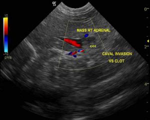
It is not possible to distinguish whether the echogenic structure invading the vena cava is clot, mass invasion, or both.

History

This 14-year-old MN DSH presented for weight loss, increased respiratory effort, and progressive anorexia. The physical exam confirmed minor tachypnea, poor body score, grade 2/6 systolic heart murmur, but was otherwise normal. The CBC presented mild leukocytosis with a left shift. The blood chemistry was normal except for a moderate elevation in amylase and mild elevation in CPK. Urinalysis: PH 6.5, USG 1.026, glucose 2+, blood 3+, epithelia rare.

Clinical Differential Diagnosis

Pancreatitis, neoplasia, cardiac disease, feline asthma, Heartworm disease, thromboembolic disease causing the respiratory embarassment.

Sonographic Differential Diagnosis

Adrenal adenocarcinoma or pheochromocytoma with caval invasion, less likely benign hyperplasia with caval clot. Lung lesion: lung metastasis, thromboembolism, infammatory consolidation, infectious disease.

Sampling

Post mortem examination revealed right adrenal undifferentiated adenocarcinoma with medullary obliteration and confirmed pulmonary metastasis after histopathological review of the lesions presented here.

Comments

The lung lesion noted was only one of many diffuse lesions found on post mortem. The radiographs presented at the time of the sonogram were from 1 week prior and were interpreted as normal with no visible lung densities noted. This was one of 3 lesions that were close enough to the thoracic wall to obtain an echogenic window without lung air artifact. The patient was euthanized due to clinical decline 7 days post diagnosis. Referring Practitioner: Stacey Lipitz-Kilcullen DVM, Little Silver Animal Hospital, Little Silver, NJ, USA.

Videos

A 2 cm right adrenal mass is seen with slight invasion into the vena cava.
Video of a 1 cm hypoechoic right middle lung consolidation adjacent to the thoracic wall with a corresponding "shower curtain" sign. This region would not be normally visible in a normally inflated lung due to air artifact.