SDEP Telecytology: Samples of cell collections; what to look for on your slides.
Have you ever gotten a "great" cytology sample from an FNA, only to find out from the pathologist that you either missed the target altogether or didn't image the correct cells on your telecytology send? It has happened to us all at one point or another. Below is a collection of still images and videos sampled from various sites to show you what you should be looking for before you send it out to the pathologist. Getting back a non-diagnostic report is the pits! :)
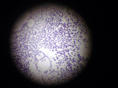
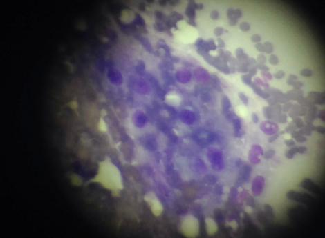
Slide on 10 x manification of a liver aspirate
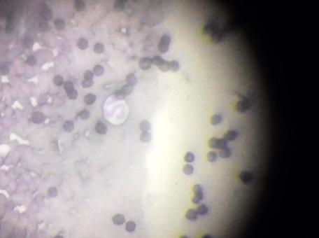
Slide on 40 x magnification
Slide on 100 x magnification with oil immersion
These all look fairly cellular right? Well yes and no, yes they have cells on them, but no they are not hepatocytes (the liver cells that needed to be evaluated).
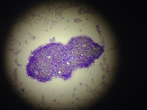
A more thorough re-scan of these same slides yielded far better results once an actual cluster of nucleated hepatocytes were recognized.
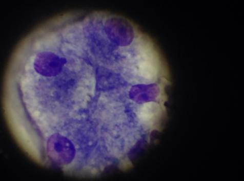
Hepatocyte cells at 10 x magnification.
Hepatocyte cells at 40 x magnification
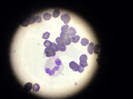
Hepatocytes at 100 x magnification with oil immersion.
Another set of hepatocytes at 100 x magnification with oil immersion.
The key to good cytology stills and video, is to find nucleated cells in the slide first, then clumps of nucleated cells if possible and scan those regions.