There was some ring formation beneath the aortic valve with the most prominent portion originating from the interventricular septum.

Image 2 and 3: Colour Doppler showed a massive turbulence across the aortic valve and a mild aortic insufficiency (Image 2 apical, Image 3 Right Parasternal Long Axis).

Right parastenal long axis view demonstrates color flow turbulence indicative of aortic insufficiency that accompanies subaortic stenosis.

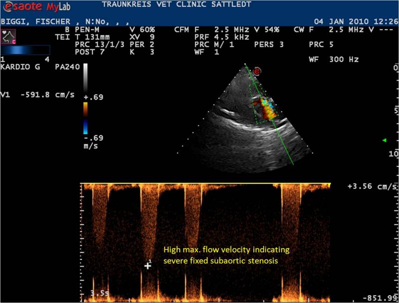
Image 4 and 5: The pressure gradient was estimated using spectral Doppler (Image 5 CW Spectral Doppler Suboptimal Angle > 15 Degrees Theta, Image 6 Optimal Angle < 15 Deg Theta). Maximal velocity was 5,92 m/s; which corresponds to a pressure gradient of 140 mm Hg. Hence, the diagnosis was high grade Subaortic Stenosis. Therapy was initiated using Atenolol at a starting dosage of 0,5 mg/kg bid and titrated weekly up to 1,5 mg/kg bid.