A 9-month old mixed breed dog was presented because of rapid fatigue during exercise and a heart murmur heard by the referring veterinarian. Clinical abnormalities: Heart murmur ejection type left heart base 5/6, heart murmur 3/6 right apex.
Image 2 and 3: Lateral & VD XRays: prominent right heart, prominent pulmonary segment. Vertebral Heart Score 10,75.
ECG: Sinus rhythm 120/min, right axis deviation.
Spectral CW Doppler placed over the color flow tirbulence over the tricuspid valve jet reveals a tricuspid insufficiency velocity of 4.5 m/sec.
Comments
The dog is scheduled to undergo balloon valvuloplasty for the pulmonic stenosis in the hopes of reducing the pressure gradient to a manageable level long term.
Videos
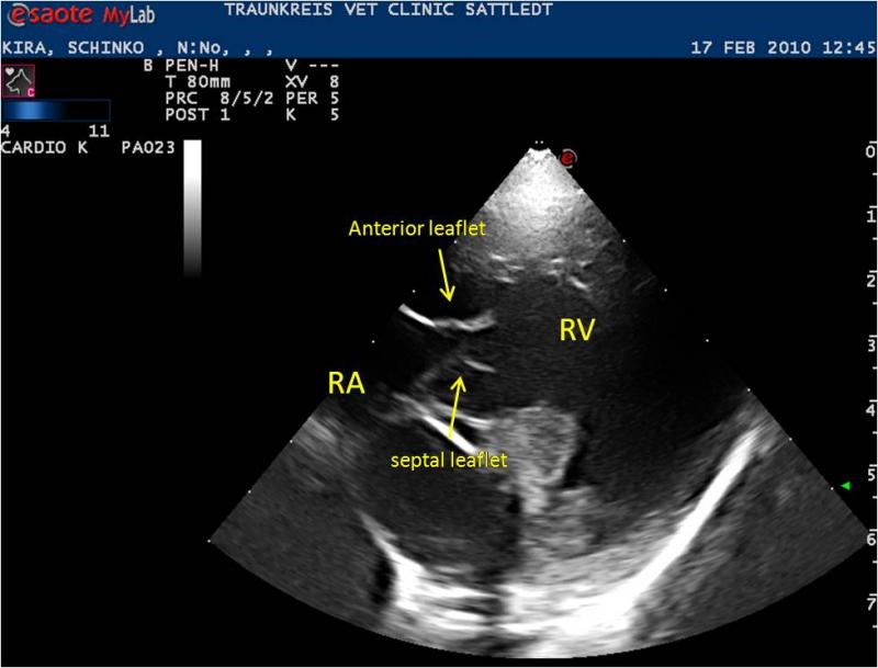
Right parasternal 4-chamber long axis ultrasound: Elongated and dysplastic tricuspid valve leaflets are evident. Color flow Doppler evidences significant regurgitation and turbulence.
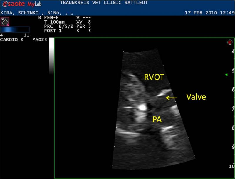
Video 2 & Image 5: Right parasternal view of the pulmonary valve demonstrating valvular doming. Pressure gradient across the pulmonary valve was 67 mm Hg, indicating moderate pulmonic stenosis.
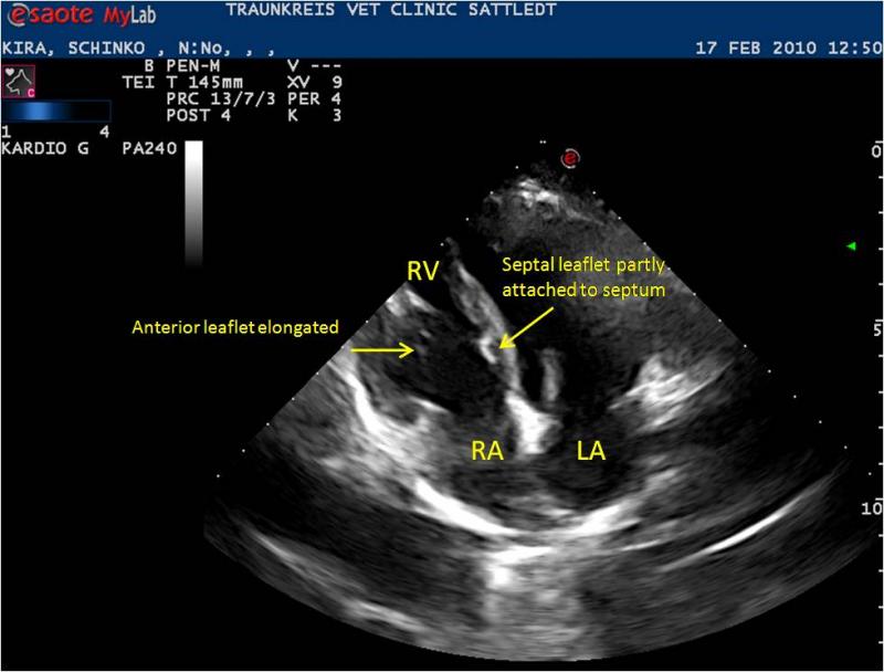
Apical 4-chamber view revealing a “tethered” septal tricuspid leaflet.
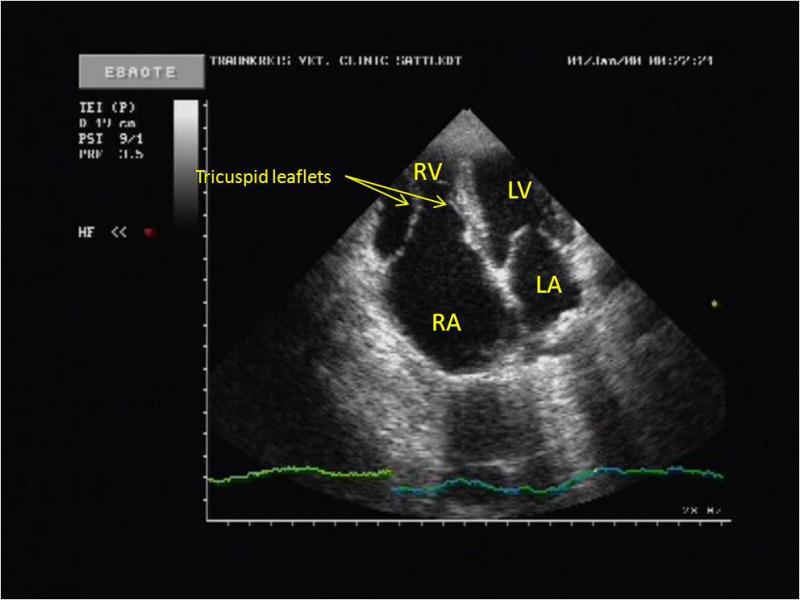
Videos 4,5 & Images 6,7: Apical 4 chamber and 5 chamber views. Hypertrophic right ventricle, dilated right atrium, dysplastic tricuspid valve (Ebstein anomaly) are all visible in these views. Severe tricuspid insufficiency is noted on color flow Doppler. This patient has Ebstein Anomaly which is defined as: a heart defect in which the tricuspid valve is abnormally formed and placed lower than normal in the right ventricle. The tricuspid valve normally has three “flaps” or leaflets. In Ebstein’s anomaly, one or two of the three leaflets are stuck to the wall of the heart and do not move normally. The valve is lower than normal in the right ventricle. Often there’s also a hole (atrial septal defect) in the wall between the heart’s two upper chambers (not present in this patient).
Videos 6,7 & Image 8: Short axis view of the tricuspid valve and elongated leaflets with eccentric right ventricular hypertrophy. Color flow Doppler demonstrates the degree of tricuspid insufficiency.


Comments