Contact Us or call 800-838-4268 for more information
Skip to main content

Endocarditis

Patient Information

Age
9 Years
Gender
Male, Neutered
Species
Feline

Images

History

A 9-year-old castrated male cat was presented for evaluation of progressive weight loss with reduced activity and appetite. Four months prior to presentation the cat had been diagnosed with severe stomatitis and dental disease that had been managed by dental scaling and extractions, which had resulted in complete resolution of the stomatitis. Three days prior to and for 10 days after the dental therapy the cat had been treated with clindamycin.

On clinical examination weight loss with moderate muscle atrophy, pyrexia (40.5° C), and a small wound on the left shoulder area were present. No abnormalities were detected on thoracic auscultation. Urine and faecal analyses were both within normal limits. Marginal non-regenerative anaemia, band neutrophilia, hypoalbuminaemia, and hyperglobulinaemia were evident on haematology and serum biochemistry. On serum protein electrophoresis the hyperglobulinaemia was quantified as marked gammaglobulinaemia and mild alpha 2-globulinaemia, indicating chronic antigenic stimulation. FIV antibody and FeLV antigen were both negative.

Further diagnostics
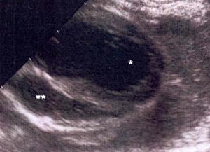
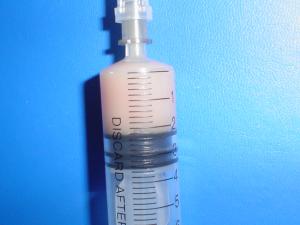
Abdominal ultrasonography was within normal limits. On a lateral survey thoracic radiograph mild border effacement of the heart and fissure lines were evident; whereas a widened caudal mediastinium was evident on a dorso-ventral survey thoracic radiograph. Right lateral echocardiography showed the presence of anechoic fluid within the pericardial sac XX on image, normal cardiac parameters, and no pleural effusion evident. The radiographic changes were ascribed to pulmonary congestion. Under ultrasound guidance approximately 20 millilitres of purulent fluid was aspirated from the pericardial sac. On cytology of the fluid the majority of the cells were degenerative neutrophils, few active macrophages were present, and there were free and phagocytosed bacteria. Aerobic and fungal cultures of the fluid were both negative; whereas a pure growth of Peptostreptococcus was identified on anaerobic culture.

Outcome

The cat was treated with amoxicillin-clavulanic acid twice a day. Within 48 hours of starting the antibiotics, the pyrexia had resolved and the cat was eating. The antibiotic therapy was continued for a further 3 weeks. On re-assessment 21 days later the cat showed marked improvement in appetite and activity. Clinically the cat showed weight gain and no pyrexia. The anaemia, band neutrophilia, and hypoalbuminaemia had resolved, and the hyperglobulinaemia had improved. Survey thoracic radiographs were within normal limits and right lateral echocardiography showed resolution of the pericardial effusion. Six weeks after the re-assessment, the owner reported telephonically that the cat was clinically well.