Fungal cultures are a common in-house lab test, but need to be performed and evaluated correctly for accurate diagnosis of ringworm.
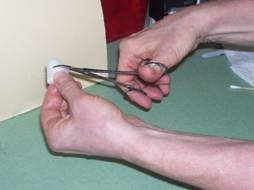
Figure 1: Clean the sampling hemostats with cotton ball moistened with alcohol. This will help prevent any introduction of bacteria into the culture which may make interpretation more difficult.
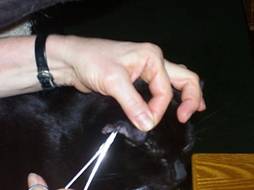
Figure 2: Use the hemostats to pluck a couple of pieces of hair from the margin of the lesion (ringworm will be actively growing at the margin). Pluck from the base of the hair. If there is no definitive lesion, you can use a new toothbrush to brush the pet all over, and then use the hemostats to place hairs from the toothbrush onto the culture medium. Yes, I should be wearing gloves; in this case I used my own kitty as a guinea pig.
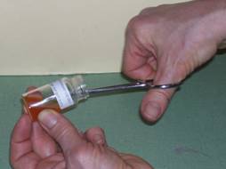
Figure 3: Place the sample carefully on the top of the medium, making sure to press gently so the hair is in contact with the medium but the medium is not disrupted.
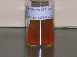
Figure 4: Apply the cap LOOSELY, label with the date and pet's name, and place in a key location where it will be checked daily. You can write the identifying information on the top of the cap.

Figure 5: Create a log such as this one. Every day for the following 10 days, the culture needs to be evaluated for presence of colony or red color, and recorded as such.
In order for a culture to be positive for ringworm, red medium color must appear first, or appear at the same time as a colony. If a colony appears first, it is likely not ringworm. Remember also that all culture media will turn red if left sitting on the shelf long enough.
To be sure it is a definitive diagnosis, you should get comfortable doing a microscopic evaluation. Place a small drop of stain on a new slide; new methylene blue is fine and most hospitals have this. Take a piece of clear tape and press it gently on top of the sample colony, then place it over the stain on the slide. Examine under 10 power for the spores of microsporum canis, trichophyton mentagrophytes and microsporum gypseum. You can generally find pictures of these in a dermatology reference book which is probably sitting on your veterinarian’s shelf.
