Clinical Differential Diagnosis
Mitral insufficiency, left sided congestive heart failure.
Image Interpretation
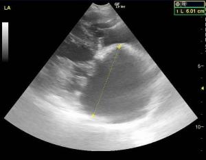
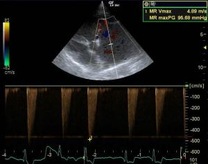
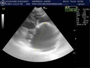
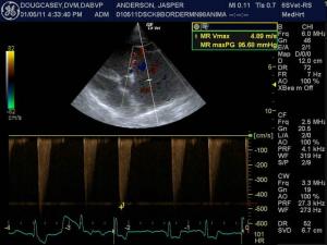
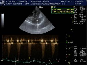
This patient presented an enlarged left atrium and LA/AO ratio was increased. The cranial and caudal mitral valve leaflets demonstrated mildly vegetative contour, lack of apposition and mild prolapse of the anterior leaflet. The left ventricle demonstrated excessive volume. Ventricular function was deemed adequate to subnormal in light of volume overload indicating potential emerging myocardial insufficiency expressed by the fractional shortening measurement of 36%. Moderate tricuspid valve insufficiency was noted at 2.6 m/sec consistent with emerging pulmonary hypertension secondary to primary left sided CHF. The right atrium was prominent.
Sonographic Differential Diagnosis
Mitral insufficiency, left atrial enlargement, early left CHF and mild early secondary pulmonary hypertension. Emerging myocardial insufficiency.
DX
Mitral insufficiency, left atrial enlargement, early left CHF, early secondary pulmonary HTN
Outcome
The patient was managed on triple therapy; furosemide, ace inhibitors and Pimobendan and stabilized over the following days and was largely asymptomatic at a 3 month recheck with minor exercise intolerance.



Comments