Clinical Differential Diagnosis
Abdominal mass - organ neoplasia (origin in spleen, liver, kidney, GI tract, ovary, adrenal, or lymph node), granuloma (mesentery, organ associated, fungal disease, or a perforated and encapsulated foreign body), intestinal obstruction (neoplasia, foreign body), hydronephrosis.
Image Interpretation
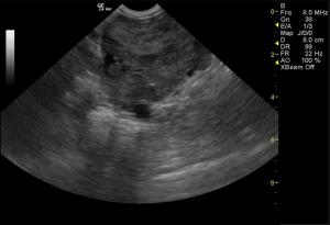
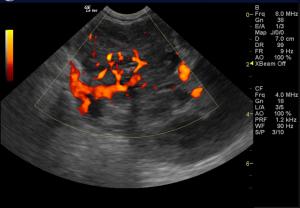
A large micronodular, dramatically hypoechoic, strongly vascular and invasive mass is present arising from the left adrenal gland, invading the phrenic vein and vena cava. Mass affect upon the left kidney is also present.
Sonographic Differential Diagnosis
Invasive left adrenal mass is seen in this study; the lack of Cushingoid clinical signs points to likely pheochromocytoma, although a nonfunctional adenocarcinoma possible.
DX
Invasive adrenal mass adneocarcinoma or pheochromocytoma
Outcome
Blood pressure measurements were recommended. Low dose dexamethasone suppression test to assess the functionality of the adrenal tumor was recommended as well, especially if urine specific gravity was less than 1.020 (UA was pending). A guarded to poor long term prognosis was given.


