Clinical Differential Diagnosis
Congenital defect - PDA, atrial septal defect (ASD), ventricular septal defect (VSD), aortic stenosis, tricuspid dysplasia.
Image Interpretation
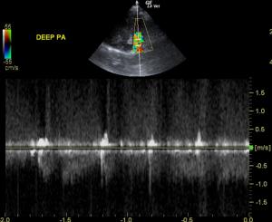
The right ventricle was of normal size (1/3 diameter of LV), echogenicity and thickness. There was no evidence of dilation or restriction. The right ventricular outflow tract was largely normal; however, the pulmonic valve, even though only slightly thickened, revealed significant insufficiency at 4.3 m/sec. Post-valvular forward flow was largely normal at 1.6 m/sec. However, the deep pulmonary artery (prior to the bifurcation) revealed a large amount of turbulence. Holosystolic flow was noted at 5 m/sec. This appears compensated at this time.
Sonographic Differential Diagnosis
Patent ductus arteriosus. Secondary pulmonic insufficiency, which appears compensated at this time.
DX
PDA, secondary pulmonic insufficiency
Outcome
The owner was considering surgery to correct the PDA at the time of last communication.


Comments