Contact Us or call 800-838-4268 for more information
Skip to main content

Acute Renal Failure

Patient Information

Age
10 Months
Gender
Male, Intact
Species
Canine

Images

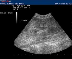
Diffuse hyperechoic cortical changes were evident with excessive cortex/medulla ratio. Focal mineralization was noted at the corticomedullary rim. Upon reviewing the images again there was no distinctive corticomedullary 'band'.
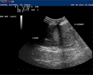
Distinct hyperechoic renal cortex is noted when comparing the right kidney to the adjacent liver. In the normal patient the renal cortex is usually isoechoic to hypoechoic when compared to the liver especially in an adolescent dog.

History

Acute Renal Failure & The Sonogram

Why are my miniature poodles dying of acute renal failure? Is it Leptospirosis? Is it a toxin? Jay Stefannacci DVM, DACVR of VCA Veterinary Emergency & Referral Associates, Norwalk, CT, USA sniffs out the underlying sono-pathology in the March, 2012, case of the month.

Sonogram (kidneys): Jaffe

History: A 10- month-old intact male toy poodle was presented for evaluation of acute onset of vomiting. Initial lab work had shown azotemia (creatinine 3.3, BUN 95), which progressively increased over 3 days ( creatinine 9, BUN 151). Severe proteinuria was present on urinalysis.

Clinical Differential Diagnosis

(Remo Lobetti PhD, DECVIM): Acute renal failure – toxins (ethylene glycol), sepsis, trauma Congenital renal disease with acute decompensation Addison’s disease

Image Interpretation

(Stefanacci VMD, DACVR):

Sonographic Differential Diagnosis

(Stefanacci VMD, DACVR): Ethylene glycol toxicosis, other toxin, infectious disease.

Sampling

Post mortem h istopathology revealed moderate, acute, diffuse nephrosis with intratubular oxalates.

Outcome

The patient was euthanized three days after presentation owing to non responsive acute renal failure.

Comments

Special thanks to Dr. Jay Stefanacci at VCA Veterinary Emergency & Referral Associates, Norwalk, CT, USA for the submission of this case.

Videos

The gastric fundus demonstrates retention of anechoic fluid and lack of peristalsis consistent with stasis/ileus and likely uremic gastritis.
Diffuse hyperechoic cortical changes were evident with excessive cortex/medulla ratio. Focal mineralization was noted at the corticomedullary rim. Upon reviewing the images again there was no distinctive corticomedullary 'band'.
Distinct hyperechoic renal cortex is noted when comparing the right kidney to the adjacent liver. In the normal patient the renal cortex is usually isoechoic to hypoechoic when compared to the liver especially in an adolescent dog.