Contact Us or call 800-838-4268 for more information
Skip to main content

Acute Splenic Torsion In An 8-Year-Old MN Shepherd Mix Canine: Our Case of the Month April 2017

Patient Information

Age
8 Years
Gender
Male, Neutered
Species
Canine

Keywords

Images

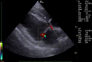
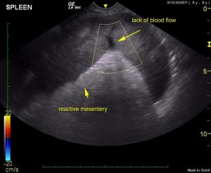
Lack of blood flow at splenic hilus on CF Doppler. Reactive mesentery. Splenic torsion confirmed.
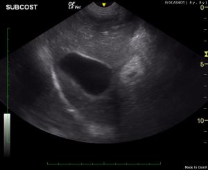
Subcostal liver and gallbladder. Note the normal parenchyma and no evidence of infiltrative disease.
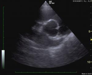
Heart base. Normal cardiac size and volume without pericardial effusion.
LA/AO June Boon measurement is normal.
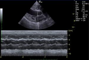
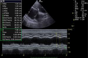
LV M-Mode. Normal cardiac function/contractility.
Mitral valve. Trivial mitral insufficiency.
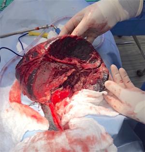
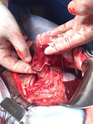
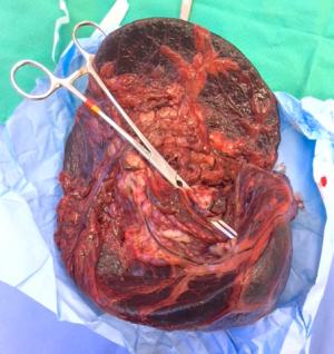
Exploratory/splenectomy confirming splenic torsion.
Splenic torsion where it should be, in and out of body experience:)

Big spleen, sudden onset, reactive mesentery & nothing else that says neoplasia? Think splenic torsion, remembering the one key button to push is the color flow Doppler over the splenic vasculature. Splenic torsions can look like splenic lymphoma sonographically, but torsions shut off the vasculature. This quick thinking clinical sonographer maneuver will save a life because you don't let the sun set on a pyometra and you don't let another minute pass on a splenic torsion. Great work by Sierra Veterinary Specialists & clinical sonographer Loetitia Saint-Jacques RVT, LVT of Paws Mobile Sonography, Lake Tahoe, CA, USA.

History

The patient was presented on emergency from his RDVM. Thoracic radiographs were unremarkable. 2-view abdominal radiographs showed a cranial abdominal mass. CBC: HCT 32%, Anemia, elevated WBC 29.58, Mono 59, Neut 22.44, PCV/TP 36%/8.2 upon presentation. A double cavity ultrasound was performed.

Image Interpretation

The spleen in this patient revealed severe enlargement and reactive mesentery. Irregular contour was noted as well as micronodular changes that extended throughout the abdomen. This is strongly consistent with splenic torsion and appeared to be isolated to the spleen. A slight amount of free fluid was noted in the caudal abdomen adjacent to the bladder. Reactive mesentery was noted throughout the mid-abdomen. The liver was slightly swollen, yet not obviously enlarged in the pathological process. Normal echocardiogram with trivial mitral insufficiency.

DX

Acute splenic torsion with regional peritonitis.

Outcome

Immediate exploratory surgery was recommended. Underlying neoplasia is possible, yet unlikely. A splenectomy was performed. No contraindication to anesthetic procedure based on echocardiogram of normal volume, function and lack of right auricular masses or pericardial effusion.

Videos

Diffusely hypoechoic swollen spleen with moth eaten appearance and hyperechoic reactive mesentery associated with the capsule. Differentials include splenic torsion, round cell or other neoplasia and less likely, splenitis.
Separate angle showing very bright echogenic mesentery associated with splenomegaly.
Color flow Doppler on the splenic vasculature is key maneuver when suspecting splenic torsion. Note the lack of signal in the splenic artery and vein confirms splenic torsion necessitating immediate surgical intervention. Ensure you turn up the gain on the CF Doppler until it speckles to see if any flow is present.
Rapid view of the right auricle (Position 3 SDEP echocardiogram manuever) is prudent with splenic pathology in case HSA is in play. In our studies we find a 15% concurrence of pericardial effusion or right auricular masses in dogs with splenic tumors.
Normal cardiac volume and contractility without contraindication for surgery.