Contact Us or call 800-838-4268 for more information
Skip to main content

Aortic Body Tumor, Mitral Insufficiency, Pulmonary Hypertension

Patient Information

Age
11 Years
Species
Canine

Images

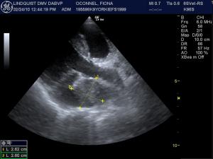
An oblique 5-chamber right parasternal view reveals an echogenic mass deriving from the post valvular aorta and superimposing on the left atrium
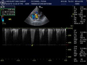
Color flow and spectral CW Doppler assessment of the mitral valve indicating severe insufficiency owing to complete color doppler filling of the left atrium and an insufficiency jet of nearly 5 m/sec. This is consistent with the clinical exam findings of a grade 4/6 systolic murmur in the region of the mitral valve. Note: Severity of MR is depemdent on the amount of filling of the LA with color flow and not on the velocity. The subnormal MR velocity was an underestimation owing to a technical issue with the probe used during the exam. The proper MR velocity should be around 6 m/sec or more owing to the systemic hypertension noted in the history.
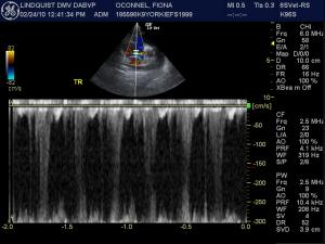
PW Doppler over the tricuspid valve in the right atrium revealing aliasing of the jet beyond 3 m/sec (Pulmonary hypertension/PH = TR jet > 2.8 m/sec) indicating a need for CW Doppler assessment to quantify the jet. From here we know that pulmonary hypertension is present but we cannot grade the degree of PH without quantifying the spectral TR jet.
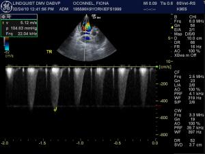
CW and Color flow Doppler assessment of the TR jet showing a clean envelope and TR velocity of 5.12 m/sec. This is consistent with severe pulmonary hypertension. Note: The pulmonary outflow velocity was 0.8 m/sec (WNL) ruling out pulmonic stenosis.

History

Aortic Body Tumor, Severe Mitral Insufficiency, Severe Pulmonary Hypertension. Sono-Pathology; 3 For 1 Deal.

Case presentation, echocardiogram performed by Eric Lindquist DMV (Italy), DABVP (Canine & Feline Practice). Founder SonoPath.com, Manager New Jersey Mobile Associates, Sparta, NJ, USA. Assessment & Tx recommendations by Dr. Peter Modler, Dipl.-Tzt. Dr.med.vet., FTA für Kleintiere (Small Animal Specialist, Austria).

History: Fiona is an 11-year-old Yorkshire Terrier that presented for exercise intolerance but was otherwise normal. A grade 4/6 left sided murmur in the region of the mitral valve was noted on clinical exam. Blood pressure was 270/155 mmHg with a heart rate of 150 BPM.

Clinical Differential Diagnosis

Cardiologist Assessment: Dr. Peter Modler, Dipl.-Tzt. Dr.med.vet., FTA für Kleintiere (Small Animal Specialist, Austria) A mass adjacent to the aortic body, most likely an aortic body tumor. Left ventricular eccentric hypertrophy due to volume overload. Left atrial dilatation. Mitral insufficiency due to mitral prolapse and degenerative changes. Tricuspidal insufficiency, Tricuspid dysplasia. Right ventricular hypertrophy Tricuspid regurgitation with a pressure gradient ~104 mm Hg Mitral regurgitation with a pressure gradient of ~96 mm Hg Combining these findings the following pathologies:

Sampling

Aortic body tumor High-grade mitral insufficiency Tricuspid valve dysplasia with moderate insufficiency Severe Pulmonary hypertension On the basis of our findings I would initiate triple therapy (Furosemide, Pimobendan, ACEI). I would probably add L-arginine (0,5-2g twice, depending on the size of the dog; L-arginine is a NO-precursor, there are no studies available on it´s use in veterinary medicine but it experimentally lowers pulmonary pressure and is – in human medicine – sometimes combined to other agents such as bosentane) and perhaps sildenafil (Viagra). Prognosis is guarded. It is questionable whether elective pericardectomy should be performed for the aortic body tumor. The median survival time following pericardectomy is 730 days, without pericardectomy it is 42 days.

Comments

Special thanks to: Dr. Erno Hollo and staff at Basking Ridge Animal Hospital, Basking Ridge, NJ, USA for providing this patient for evaluation.

Videos

Live view of the same mass demonstrating deviation of the aorta by the mass. Slight mitral valve prolapse is noted with mild-moderate left atrial enlargement.
Short axis heart based view demonstrating the aortic body tumor, pulmonary outflow tract to the right and left atrium and tricuspid valve to the left. Slight clubbing and vegetative changes are noted on the tricuspid valve.
Oblique apical view demonstrating the full width of the aortic body mass an maximizing the left atrial enlargement (nearly 5 cm) that is consistent with chronic dilation and rotated out of the typical la/ao view likely owing to the mass deviation.