Contact Us or call 800-838-4268 for more information
Skip to main content

Aortic Dissection In A 16-Year-Old MN DSH Cat: Our Case Of the Month November 2022

Patient Information

Age
16 Years
Gender
Male, Neutered
Species
Feline

Images

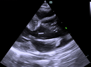
5-chamber long axis view of aortic dissection.
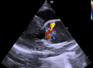
Color Doppler over aortic dissection.
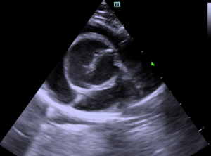
Short axis heart base view of aortic dissection.

"Aortic dissection (AD) is characterized by bleeding within the aortic wall or a tear in the intimal layer of the aortic wall, resulting in the passage of blood from the aortic lumen into the tunica media. In cases of AD, a floating, intimal flap in the aortic lumen divides the lumen into a true portion, with flow present, and a false portion, with no flow." Exerpt from Aortic dissection in four cats: clinopathological correlations. Oricco/Perego/Poggi/Tursi/Biasato/Santilli. Click this link to hear from Dr. Peter Modler on Aortic Dissection via 5 Sono-Minutes with SonoPath.

Shari Reffi, CVT, SDEP® Certified clinical sonographer from our SonoPath Mobile Veterinary Ultrasound team, put her efficient SDEP® skills to the test with this very critical patient. STAT interpretation by Eric Lindquist, DMV, DABVP, Cert. IVUSS.

History

The patient with a history of asthma (on Flovent® and prednisolone), presented for an abdominal ultrasound due to persistently elevated liver enzymes since 3/2022, and losing weight despite increased appetite. CBC/Chem: AL:T 510, ALP 329, T. bili 1.4, leukocytosis with neutrophilia and monocytosis. Current meds: Denamarin 90 mgs q24 hrs, Metronidazole 50mgs q12 hrs, Amoxicillin 50 mgs q12 hrs. At the the time of ultrasound the patient was dyspneic and an SDEP® emergency echo was performed to assess the heart. Blood pressure post scan (tail) was 118/98 (99); patient was put in O2 chamber for support.

Image Interpretation

The aorta in this patient was dilated with a splitting cranial wall and echogenic debris, consistent with aortic dissection. The aortic valve was mildly thickened. Periodic arrythmia was present.

DX

Aortic dissection.

Outcome

Aortic dissection is a severe consequence to systemic hypertension or underlying disease process. Prognosis is poor. The causes of abdominal evaluation for cause of underlying disease is indicated with search for causes of hypertension. However, prognosis is poor, and this patient is at high risk for sudden death. Thromboembolic episodes may also be playing a role given the increased respiratory effort. EKG is indicated to assess periodic arrhythmia yet was paroxysmal during the exam.

Videos

SDEP® position, 1 long axis; aortic dissection.
SDEP® position, 2 short axis heart base.
Color Doppler across aortic dissection.