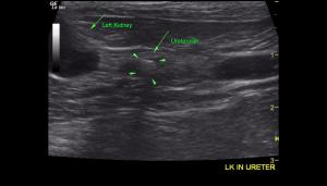
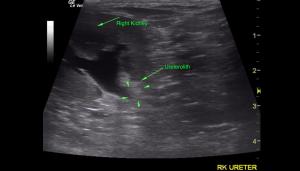
"OUCH!" Acute pain on palpation…. who would have thought there would be not one, but TWO ureteroliths causing pain in this DLH feline patient. Normal ureters are generally NOT visible on a scan, but acute pain in the region of the kidneys should clue you in to pay particular attention to the kidney region, including use of not just the micro convex probe but also the linear probe to highlight and create those “artistic” views. When you find pathology, such as a dilated ureter, FOLLOW it and see where it goes and what may be hiding there…. Our 17-point SDEP Abdomen protocol teaches you how to do this routinely with EVERY patient, and every potential pathology. Many thanks to Dr. Candace Remcho of West Hills Animal Hospital in Corvallis, Oregon for the proficient case management of this patient, as well as the comprehensive interpretation of this case by the newest addition to the Sonopath Team of Specialists, Dr. R.McKenzie “Mac” Daniel, DVM, DABVP (Canine and Feline). Also, a loud shout out to Heidi Putnam, clinical SDEP sonographer of Animal Sounds NW Veterinary Mobile Ultrasound for the exemplary imaging.
DX
Bilateral ureterolithiasis present in the right and left ureters with secondary proximal ureter dilation. This is consistent with partial to complete obstruction. Marked hydronephrosis with loss of normal renal medulla parenchyma in the left kidney and merging to mild hydronephrosis with mild loss of normal renal medulla parenchyma of the right kidney. Focal nephrolithiasis was present within the dilated renal pelvis and medulla of the right kidney.
Outcome
The bilateral ureteroliths are likely causing partial to complete obstruction of the right and left ureter with hydronephrosis in the right and left kidney. Smaller left kidney size with marked hydronephrosis suggests more chronic disease in the left kidney. The functionality of the left kidney is highly questionable. The pain exhibited by the patient is likely associated with the ureterolithiasis and development of hydronephrosis. Treatment options would likely be centered around preserving functionality of the right kidney. Referral in this case for possible surgical options such as ureterolith removal or possible stent placement is recommended. Placement of a subcutaneous ureteral bypass device (SUB) is also an option in this case. If referral is not an option then conservative treatment may include continued IV fluids, pain medications and an Alpha blocker such as Prazosin to try to get the ureteroliths to move. This may or may not be possible. Therefore, guarded prognosis is warranted in this case. Systemic blood pressure may also be considered due to the degree of renal disease.Unfortunately, due to quality of life concerns and a poor prognosis the patient was humanely euthanized.


