Contact Us or call 800-838-4268 for more information
Skip to main content

Bowel Infarction

Patient Information

Age
7 Years
Gender
Male, Neutered
Species
Canine

Images

History

Spontaneous Bowel Infarction in a Golden Retriever

History: This 7-year-old MN Golden Retriever presented for anorexia of sudden onset. The physical exam revealed fever of unknown origin, tacky mucosal membranes, and focal splinting abdominal pain cranial to the urinary bladder. The CBC revealed mild/moderate neutrophilic leukocytosis (23,000 wbc/hpf). Chemistry panel and urinalysis were uneventful.

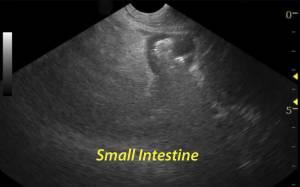
The abdominal sonogram revealed a focal painful region of distal small intestinal pathology with ill-defined hyperechoic adhesion pattern of surrounding mesentery that is attached and associated with the compromised intestine cranial to the urinary bladder. There is a complete loss of intestinal mural detail in the affected small intestine with gas penetration into the wall. The patient was extremely painful on imaging this structure. Primary differentials included perforating intestine due to underlying neoplasia (LSA, MCT, carcinoma), transmural inflammatory disease, bowel infarction, foreign body passage.

DX

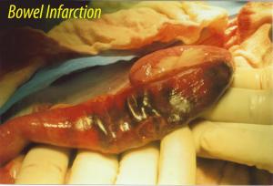
Spontaneous bowel infarction and localized peritonitis due to inflammatory bowel disease with transmural penetration.

Outcome

After bowel resection was perfomed upon immediate laparotomy, this image was taken of the compromised portion of small intestine. Histopathology: Lymphoplasmacytic enteritis, vasculitis, thrombosis, and localized peritonitis. The patient responded intitially, had a clinical setback 5 days post surgery with inflammatory peritonitis that resolved with medical therapy over the next number of days. The patient was clinically normal 3 months post surgery.

Comments

Referring Hostpital: North Jersey Veterinary Hospital

Videos