Contact Us or call 800-838-4268 for more information
Skip to main content

Chronic Cholangiohepatitis in a 1-year-old FS Australian Shepherd Mix: Our Case of The Month June 2018

Patient Information

Age
1 Year
Gender
Female, Spayed
Species
Canine

Keywords

Clinical Signs

Special Testing

Blood Chemistry

Images

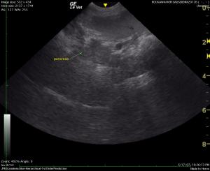
Edematous pancreas.
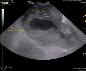
Hypoechoic liver, double-walled gallbladder, and free fluid.
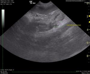
48 hours post treatment -Largely resolved pancreatitis.
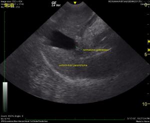
48 hours post treatment-Normal gallbladder, uniform parenchyma.

All we know is "If it’s sick it needs a probe" and if you’re treating, it needs a follow-up probe. Who knows what this young dog got into?

Presented for acute onset of collapse with hepatomegaly on radiographs, this dog was scheduled for immediate ultrasound. Given a guarded prognosis, the recommended treatment protocol was followed and a repeat scan performed 48 hours later revealed dramatic improvement and markedly less pain.

Thanks to the quick response and dedicated care of Dr. Maniar at Rockaway Animal Hospital, this young dog didn’t sit in a cage for 24-48 hours with no direction; instead after an initial ultrasound by SonoPath Mobile's own Diane McFadden, RVT, SDEP™ certified clinical sonographer, 48 hours later she was well on the road to recovery and ready for outpatient treatment. The patient is reported to be doing great following her treatments.

History

A 1-year-old, FS, Australian Shepherd mixed breed canine was presented for acute onset of collapse/dizziness. This was the first episode that the patient ever had; patient was BAR prior. Hepatomegaly was seen on radiographs. Blood chemistry showed an elevated ALT, CBC/UA were WNL. 4DX heartworm test was positive, Lepto test was negative.

Image Interpretation

The liver was swollen and hypoechoic with a double layered gallbladder. The liver revealed coarse
architecture with increased portal markings. This is consistent with acute on chronic cholangiohepatitis.

DX

Chronic cholangiohepatitis and free fluid. Pancreatic edema.

Outcome

The pancreatic edema and free fluid was likely owing to portal hypertension. Liver biopsy was suggested in this case. Treatment for acute on chronic cholangiohepatitis with coverage for Leptospirosis was recommended even though the patient is negative. Copper storage was a possibility. A very guarded prognosis was given. A recheck ultrasound was performed 2 days later which revealed structurally resolved cholangiohepatitis, normalized gallbladder, and largely resolved pancreatitis. The presentation was dramatically improved compared to the prior sonogram. Outpatient therapy was recommended as long as the patient was stable. The patient was treated with I.V. fluids, metronidazole, ampicillin, and Pepcid followed by heartworm treatment protocol of prednisone and doxycycline. The patient is reported to be doing great following her treatments.

Videos

Video demonstrating left base and left limb of the pancreas with mixed hypoechoic areas consistent with necrosis or suppurative changes and edema. Hyperechoic ill-defined surrounding fat suggests inflammation and peritonitis. Patient exhibited a + Murphy sign in this area.
The liver is coarse in architecture with increased portal markings with a thickened echogenic gallbladder consistent with cholangiohepatitis with surrounding free fluid owing to portal hypertension or inflammation.
Two day follow-up image of the same pancreas as video 1 demonstrating a well organized hypoechoic area of the pancreas; patient was no longer painful. The ill-defined hyperechoic fat pattern has completely resolved, indicative of resolving inflammation.
Liver and gallbladder essentially normalized with gallbladder no longer thickened.