Contact Us or call 800-838-4268 for more information
Skip to main content

Chronic, Focally Extensive Transmural Enteritis in an 11-Year-Old FS Tibetan Terrier: Our Case Of the Month February 2019

Patient Information

Age
11 Years
Gender
Female, Spayed
Species
Canine

Keywords

Clinical Signs

Images

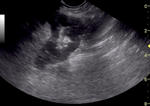
caudal abdominal intestinal mass
note loss of mural detail

Watch out for imposters...

If you hear hoof beats look for horses. If the FNA is inconclusive look for unicorns. When everything points to intestinal lymphosarcoma from a clinical and sonographic presentation, remember there are other more treatable imposters out there. This is one of those cases. 

Many thanks to Dr. John Ammeraal from American Animal Hospital for the management of this case and providing the diagnostic images. Another thanks goes out to Dr. Wendy Turnerat Andover Animal Hospital for her surgical management of this case. A joint effort with a great outcome.

History

An 11-year-old FS Tibetan terrier was presented for vomiting for 2 weeks. Physical examination found a palpable mass in the abdomen and pale mucous membranes.

Image Interpretation

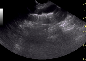
A caudal abdominal intestinal mass was noted with loss of mural detail, measuring 2.0 cm x approximately 8.0 cm, strongly suggestive for intestinal lymphoma, possibly resectable.

Sampling

Tru-cut biopsy of the intestinal mass was obtained via exploratory surgery.

DX

Focal mucosal erosion/ulceration and focally extensive transmural predominantly chronic inflammation with fibroplasia/fibrosis and serositis/peritonitis and increased mucosal eosinophils.

Outcome

Intraoperative ultrasound with resection and anastomosis is recommended or ultrasound guided FNA. This appears to be an isolated lesion. Blood loss from the intestinal mass is possible if the anemia is regenerative. Otherwise, bone marrow disease should be considered as a potential. U/S-guided FNA of the intestinal mass was performed and findings were: epithelial proliferation. The patient underwent exploratory surgery with resection and anastamosis of the affected area of intestines. During enterotomy a small (approx. 10mm x 6mm) piece of what appeared to be rawhide was found directly upstream from the mass. Biopsy of the intestinal mass found in the small intestine: focal mucosal erosion/ulceration and focally extensive transmural predominantly chronic inflammation with fibroplasia/fibrosis and serositis/peritonitis and increased mucosal eosinophils. The patient recovered from the procedure without event. The patient was found to be 100% improved at her follow up visit and owner reports she is behaving better than she has been in a year.

Videos

caudal abdominal intestinal mass, appears isolated
mass measures approximately 2 cm x 8 cm