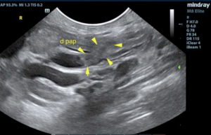
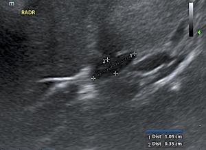
Teamwork makes the dreamwork! Team scanning is a fantastic way to learn other's imaging techniques, tips, and tricks. In this Case Of the Month, we are focused on "old cat tubes", imaging of the common bile duct, duodenal papilla, and biliary tree. This 13-year-old cat had some pathology plugging up the tubes. See what our team found with images provided by Shari Reffi, CVT, SDEP® Certified clinical sonographer for SonoPath Mobile Veterinary Ultrasound and Jessica Miller, BS, RDMS, clinical sonographer for SonoPath Mobile Veterinary Ultrasound. Interpretation of ultrasound images by Eric Lindquist, DMV, DABVP, Cert. IVUSS and cytology interpretation by L.D. McGill, DVM, Ph.D., DACVP.
Sampling
Ultrasound guided FNAs of both the spleen and liver were performed without complication.
DX
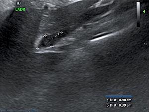
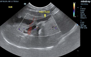
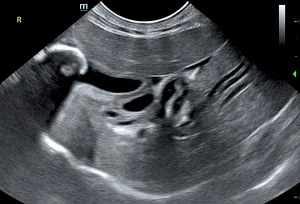
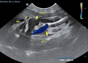
Hepatic swelling with common bile duct dilation and free fluid. Biliary calculi, may be incidental. Scalloping spleen.
Outcome
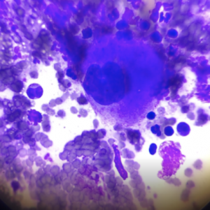
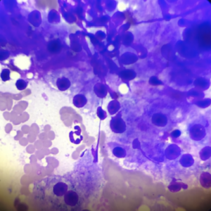
Concern for post hepatic obstruction; possibility of a concurrent hepatic lymphoma as the cause due to the clinical profile. The common bile duct appears to taper into the duodenal papilla adequately. Recommend screening via ultrasound-guided FNA of the liver and spleen to ensure that no obvious lymphoma is present followed by surgical intervention with cholecystectomy and common bile duct lavage. Ultrasound guided FNAs of both the spleen and liver were performed without complication. Cytology results: Liver - Chronic pyogranulomatous hepatitis with hepatocellular vacuolization and probable early feline fatty liver syndrome. Spleen - Chronic suppurative splenitis with extramedullary hematopoiesis.Two separate issues are likely at play in this patient with the possibility of hepatic lymphoma. At last recheck, the patient was found to be doing well on current treatments of Denamarin, Metronidazole, and Ursodiol.




Comments
Additional cytology notes: "The changes in the liver support chronic inflammation and evidence of secondary hepatocellular change. This could be due to biliary obstruction or possibly due to pancreatitis ascending up the biliary tree with inflammation. There is no suggestion of malignancy nor do we find changes supporting sepsis. The changes are more characteristic of chronic inflammation which is likely secondary to the biliary problem or pancreatitis. The changes in the spleen are likely secondary to the problems in the liver. This could be due to inflammation in the biliary tree, pancreatitis or other changes. There is no suggestion of neoplasia or sepsis in the spleen. Further treatment for the gallbladder obstruction and possible pancreatitis was encouraged in this case." - Larry McGill, DVM, PH.D., DACVP.
If you or your team are interested in SonoPath's Telecytology Services click HERE for additional information.