Contact Us or call 800-838-4268 for more information
Skip to main content

Confirmed Pyometra in an 11-year-old MN Poodle.... YES a MN!: Our Case Of the Month February 2016

Patient Information

Age
11 Years
Gender
Male, Neutered
Species
Canine

Keywords

Images

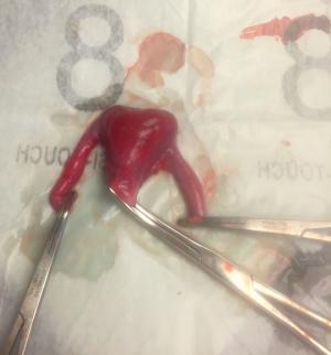
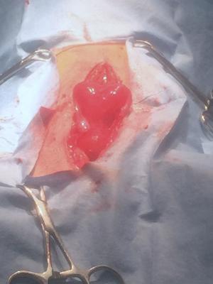
Surgical view of the hermaphroditic uterus at surgery.
Post-op pyometra in this MN miniature poodle.
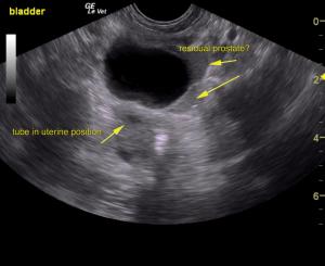
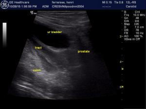
Dilated fluid filled structure in position of the uterus. Regional ill-defined fatty inflammation noted associated with the structure.The normal residual fatty prostate is seen given that this patient had been neutered. Pyometra suspected.
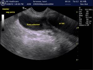
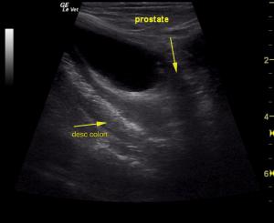
Echogenic fluid dilation of the suspected pyometra. Urethra visible and nsf.
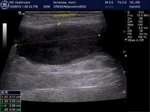
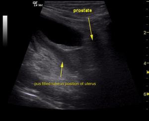
Echogenic fluid dilation of the suspected pyometra.
Identification of the echogenic dirty shadow of the normal descending colon as well as a normal urinary bladder and ureteral papillae, leads the sonographer to decide the only structure this dilated tube present between the bladder and colon can only represent a hermaphroditic uterus with pyometra.
see above
see above

What pathology lies inside this "Ain’t Doin’ Right" 11-year-old MN Miniature Poodle?… Pancreatitis, neoplasia, back pain..?? Tag team Andi Parkinson, BS, RDMS/Rachel Brilhardt, RDMS of Intrapet Imaging & Lindquist, DABVP remotely with Sonopath.com telemedicine and the art of sonographic deductive reasoning discovers a Hermaphroditic Pyometra in this Male Neutered Poodle with the organs Henri never thought he had in the SonoPath.com February, 2016 case of the month.

History

The patient was present for "ADR", back pain, pollakiuria and dysuria, and dark urine. Treatments included IVF, Ampicillin, and Baytril without clincial response. Urine specific gravity was 1.034, pH 7, 1+ protein. Bloodwork was unremarkable.

Clinical Differential Diagnosis

Orthopedic pain, urethritis, prostatic tumor, neoplasia, abscess, pancreatitis, open.

Image Interpretation

The urinary bladder itself presented minor thickening and minor debris. A tubular structure was noted in a position between the colon and the urinary bladder with dilation. Given the position of the structure and echogenic fluid dilation pyometra owing to hermaphraditism is suspected.

Sonographic Differential Diagnosis

Pyometra owing to hermaphroditism.

Sampling

Surgical removal of the infected uterus was performed confirming the suspicion of pyometra.

DX

Hermaphroditic Pyometra.

Outcome

Exploratory surgery with removal of this structure was recommended and performed. The patient responded well to surgery.

Comments

Special thanks to Rachel Brilhardt RDMS & Andi Parkisnon RDMS of Intrapet Veterinary Imaging, Baltimore, MD, USA for imaging Henri and to Dr. David Cullum & staff at Creswell Vet Clinic, Baltimore, MD, USA (www.creswellvetclinic.com) for the medical and surgical management ensuring a positive outcome.

Videos

Efficiency video long axis scan of the urinary bladder, pelvic urethra sweeping dorsally through the tubular uterus with echogenic fluid and then to the descending colon. It is important in this case to identify the normal ureteral papillae to ensure the tubes do not represent dilated ureters which would be a more common pathology in this region. Regional fatty inflammation is present. Diagnosis: Hermaphroditic pyometra