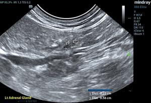
Conn's syndrome in cats is not a pathology we see every day, but an SDEP-trained sonographer is always prepared, having been drilled in the 17-point abdominal protocol including every adrenal every time, for both dogs and cats. Melding the clinical signs, labwork, and thorough ultrasound imaging allowed for a prompt diagnosis and internist referral visit within 24 hours.
Dr. Cathy Jarrett, SDEP® Certified clinical sonographer and owner of Potomac Mobile Veterinary Ultrasound (Ashburn, VA) imaged this patient, and Dr. Gouri Krishna, DVM from Silver Spring Animal Hospital managed the case. Thank you to Amy Roth Jones, DVM, DACVR for her detailed interpretation of these diagnostic images.
DX
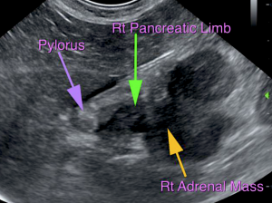
Right adrenal mass with reactive retroperitoneal fat - Suspect pancreatitis of the right pancreatic limb secondary to regional inflammation.
Outcome
This study confirmed the presence of a right adrenal mass consistent with suspected Conn’s syndrome and hyperaldosteronism. The mass appeared fairly well encapsulated and surgically resectable. Given the size, it does efface the pancreatic limb in the region and is likely causing pancreatitis. A metastatic lesion or adhesion of the mass to the pancreas is thought less likely given the separation from the retroperitoneal cavity; however, this case may benefit from a CT for a more definitive and anatomic evaluation, and assessment of the regional vasculature as well as any possible involvement of the pancreas or other surrounding soft tissues. Alternatively, exploratory laparotomy for adrenalectomy with concurrent evaluation of the pancreas is and option. For any possible involvement, a partial pancreatectomy could also be considered. Consultation with an internal medicine specialist and surgeon was recommended. After discovery of the right adrenal mass the patient was immediately referred to an internist and was seen less than 24 hours later for further discussion of the right adrenal tumor and hypokalemia. Radiographs showed no evidence of metastatic disease. Surgical excision with hospitalization was discussed with fluid therapy and potassium supplementation (optional CT per our surgeon). Supportive care and medical management with oral potassium supplementation (consider spironolactone) was also discussed. Ultimately, the owner elected humane euthanasia in this case as the patient's quality of life had declined significantly.


