Contact Us or call 800-838-4268 for more information
Skip to main content

Cranial Abdomen Mass

Patient Information

Age
16 Years
Gender
Female, Spayed
Species
Feline

Images

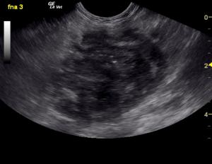
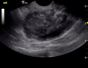
Pancreatic mass. A mixed echogenic complex and expansive mass is present in the left pancreatic base. Note the echogenic capsule is still; uniform but significantly swollen in contour.
FNA of the left pancreatic mass.

"Well that's not supposed to be there"…. mass in the left cranial abdomen. The sonogram defines if the lesion includes "crucial parts" or "extra parts" to remove. Define the lesion with a needle and remove it with a scalpel. Dx efficiency at its finest with unfortunately an inopportune outcome. Rachel Brilhart, RDMS of Intrapet Imaging in Baltimore, MD defines this pancreatic mass prior to surgery.

History

A 16-year-old FS DSH cat with history of diabetes and hyperthyroidism was presented due to vomiting, diarrhea, and anorexia. Physical exam found poor body condition and weight loss. Urinalysis showed SG of 1.017, proteinuria, and hematuria. A coagulation panel was mildly elevated.

Clinical Differential Diagnosis

Pancreas clinical signs- pancreatitis, neoplasia, abscessation. Uncontrolled hyperthyroidism. G.I. clinical signs- IBD, neoplasia, intussusception, foreign body, ulceration, infectious (viral/bacterial/parasites). Renal clinical signs- renoliths, neoplasia. Interstitial cystitis.

Image Interpretation

A left, caudal pancreatic mass was noted in this patient and measured 4.0 cm with moderate complexity attached to the left lobe of the pancreas. Hyperechoic surrounding fat was also noted. This is consistent with extension of the neoplastic process or associated inflammation. This appears potentially resectable. An exploratory surgery is recommended with removal of the adjacent lymph node measuring 1.5 cm (Not seen in these images). The right limb of the pancreas was mildly heterogenous, yet unremarkable. There was no overt evidence of metastatic disease noted. FNA was obtained of the pancreatic mass. The pancreas in this patient presented severely dilated duct at 0.49 cm with heterogenous, irregular parenchymal changes and tortuous pancreatic duct. The caudal abdomen revealed a cystic or irregular lymph node that measured 1.48 x 1.15 cm. This is likely jejunal lymph node with distorted architecture. Granulomatous disease or possible neoplasia may be present. FNA was recommended.

Sonographic Differential Diagnosis

Pancreatic mass, left limb. This appears resectable. Minor adjacent lymphadenopathy.

Sampling

US-guided FNA of the pancreatic mass.

DX

Poorly differentiated adenocarcinoma. (Spleen and adhered omentum with no recognizable pancreatic tissue present).

Outcome

Clinical recommendations included chest radiographs, assessment of the cytology followed by exploratory surgery with aggressive left pancreatectomy as well as exploration for any evidence of metastatic disease that was not noted on ultrasound. However, no metastatic disease was overtly suspected and the lesion appeared to be largely isolated to the left pancreatic limb with minor capsular escape. Pancreatic carcinoma was the primary differential, however a granulomatous lesion was also a possibility from a sonographic aspect though less likely. The patient underwent exploratory surgery. The lesion was resected surgically even though it was wrapped around the splenic vessels. A concurrent splenectomy was also performed. The patient unfortunately died suddenly post-op after succesful resection. Thromboembolic episode was suspected.

Comments

We at SonoPath.com would like to thank Andi Parknson RDMS & Rachel Brillhart RDMS of Intrapet Imaging, Baltimore, MD and their clients and collaborators, Dr. Alicia McMichaels (primary) and Dr. Jonathan Kaufman of Eastern Animal Hospital, Baltimore, MD, USA (http://easternanimalhospital.com/) for their excellent clinical and surgical management of this case. Moreover, their continual support of diagnostic efficiency in veterinary medicine through direct and remote support in their operations in exemplary and paramount to success in this field. Even though the outcome was not a positive one in this patient, the diagnostic workflow allowed the owner to make solid and efficient decisions for their pet with minimal suffering or delay in the diagnostic process in defining this aggressive neoplastic pathology.

Videos

Complex mixed echogenic left pancreatic mass expanding medially and displacing the portal vein (color flow) and the right pancreatic limb out of view.
Pancreatic mass in the midst of surrounding omentum in the cranial abdomen. Note the ill defined fat associated with the expanding capsule which indicates associated regional inflammation. Capsular escape is starting to occur ion the near field toward the end of the video clip.
Note the stomach and pyloric outflow with the adjacent right pancreatic base and right limb of the pancreas (1-2 cm of depth) that is not affected by the mass and is slightly hypoechoic to surrounding fat typical of older feline pancreas with potential concurrent inflammation but obviously not infiltrated with neoplasia. Not how the mass is linked to the dilated pancreatic duct which indicates the origin of the mass. This efficiency clip of the pancreas was key to definitively identify the mass origin as pancreatic.
FNA of pancreatic mass. Note that the needle is clearly visible in its entirety and traverses variable echogenic changes which is key to obtaining a variety of cells within the lesion and to identify the cytology as pancreatic in origin. Often neoplasia can be undifferentiated or be present along with necrosis and inflammation. Therefore. a sample of a single echogenic change will minimize the potential for a complete diagnosis including in this case pancreatic cells, adenocarcinoma, and inflammation.