Contact Us or call 800-838-4268 for more information
Skip to main content

Cyanosis

Patient Information

Age
6 Years
Gender
Female, Spayed
Species
Canine

Images

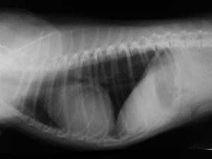
(Radiographs): generalized right sided cardiomegaly is noted. There is no sign of pulmonary congestion visible. The lung pattern is unremarkable
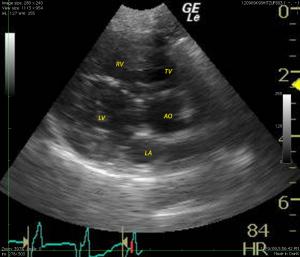
(Ultrasound): The cardiac presentation in this patient presented normal to mildly subnormal left atrial size. Normal apposition of the mitral valve was present with age related changes. The left ventricle had concentric hypertrophy/pseudohypertrophy primarily at the left ventricular septum with hypovolemia.
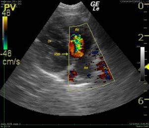
The aortic valve was mildly thickened with a small membranous VSD noted prior to the AV. Color flow Doppler passage left to right was noted through the VSD on apical view. The right ventricle was significantly thickened in this patient. (Not shown) The pulmonic valve appeared mildly thickened. Pulmonic outflow velocity was mildly elevated at approximately 2.3 M/S. Post valvular pulmonic artery was dilated. Pulmonic insufficiency was evident but not able to be adequately quantified. Clinically significant pulmonic stenosis does not appear present in this case.
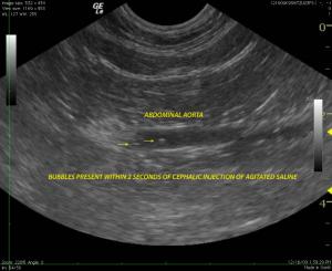
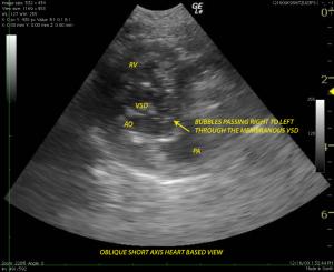
A bubble study was performed with agitated saline injected into the cephalic vein. Images of the abdominal aorta revealed bubble passage within 2 seconds of injection indicating venous aortic shunting. Four chamber long axis view of the heart also revealed passage of bubbles from the right ventricle into the aortic outflow through a small membranous VSD
Bubbles injected in the cephalic vein reach the right atrium and tricuspid valve demonstrate right to left shunting through the VSD as they enter into the aortic outflow (Mercedes benz sign) from the right ventricle which, in turn, accepts the majority of the bubbles.

History

"Tiny Bubbles" & veterinary medicine from the inside out:
Reverse PDA & Reverse VSD evidenced by a bubble study comprises the July, 2011 SonoPath case of the month. The patient was referred by Penninsula VH & imaging performed by English Bay Ultrasound Service; Doug Casey DVM, DABVP, Vancouver, B.C. Canada. Interpretation & support by Lindquist & Modler of SonoPath.com

Sonogram: Fadara examined at Penninsula VH. Imaging performed by English Bay Ultrasound Service; Doug Casey DVM, DABVP, Vancouver, B.C. Canada.

History: A 6-year-old FS Shiht-Zu Dog presented for exercise intolerance and cyanosis under stress. The clinical exam was otherwise unremarkable. A slight right and left sided murmur was auscultated. Right sided cardiomegaly was noted on radiographs.

Clinical Differential Diagnosis

(Modler FTA für Kleintiere – Cardiology): Differentials for heart murmur and cyanosis of cardiac origin are: VSD with Eisenmenger´s syndrome (R-L shunt) VSD with bidirectional shunt PDA with bidirectional shunt PDA with R-L shunt VSD combined with pulmonic stenosis Tetralogy of Fallot Differentials for heart murmur and cyanosis due to pulmonary disesase are: Pulmonary pathology causing pulmonary hypertension with tricuspid and concomitant mitral regurgitation (small breed dog)

Image Interpretation

(Lindquist DMV, DABVP and Modler FTA fur Kleintiere - Cardiology)

Sonographic Differential Diagnosis

Right ventricular hypertrophy, reverse PDA/AP window. Small membranous ventricular septal defect with bidirectional flow.

Outcome

The patient was referred to a university cardiologist and passed away prior to potentially high risk interventional therapy could be attempted.

Comments

(Modler): Dogs can sometimes have a cyanotic tongue during exercise without having cardiac or circulatory compromise. This can simply be found out by measuring the PCV (is elevated in patients with R-L-shunt). There is a general rule that covers about 90% of exercise intolerance cases with cyanosis: Young cyanotic dogs usually have a cardiocirculatory shunt from R to L Middle aged dogs can have either point 1. Or 3. Old cyanotic dogs usually have pulmonary disease (can also be pulmonary edema) Cyanosis that is noted only on the tongue and occurs during exercise, not causing collapse or breakdown, is most likely not of cardiac origin. Dogs obviously have the possibility to recirculate blood within the tongue thus extracting all the oxygen from the blood. This cyanosis is not noted on the conjunctival membranes or gingival and lasts only during exercise. Note that dogs having a R-L shunt do not always have a noticeable heart murmur! Sometimes it is inconstant and very slight - or - if there is a very large shunt with R-L shunt there is no heart murmur present.

Videos

Bubbles injected in the cephalic vein reach the right atrium and tricuspid valve demonstrate right to left shunting through the VSD as they enter into the aortic outflow (Mercedes benz sign) from the right ventricle which, in turn, accepts the majority of the bubbles.
The aortic valve was mildly thickened with a small membranous VSD noted prior to the AV. Color flow Doppler passage left to right was noted through the VSD on apical view. The right ventricle was significantly thickened in this patient.
Bubbles injected in the cephalic vein reach the right atrium and tricuspid valve demonstrate right to left shunting through the VSD as they enter into the aortic outflow (Mercedes benz sign) from the right ventricle which, in turn, accepts the majority of the bubbles.
Heart based view revealed bubble passage also from the pulmonary artery into the aorta at the level of a PDA or arterial-pulmonary window. This would indicate venous to aortic shunting through a reverse PDA/AP window and a small revering VSD consistent with Eisenmenger’s physiology. However this should be confirmed with alternate views or fluoroscopy. The VSD demonstrated bidirectional flow with the predominance on color flow as left to right and minor reversal right to left during the bubble study.