Contact Us or call 800-838-4268 for more information
Skip to main content

Disseminated lymphoma in an 11-year-old FS DSH cat: Our Case Of The Month January 2012

Patient Information

Age
11 Years
Gender
Female, Spayed
Species
Feline

Keywords

Images

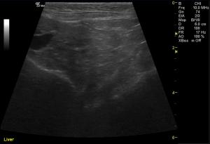
The liver was moderately enlarged, rounded in shape having mildly hypoechoic echogenicity with multifocal hypoechoic and cystic nodules throughout. The gallbladder was normal in size, shape, and clean.
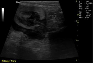
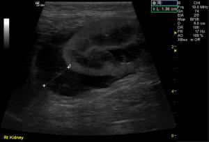
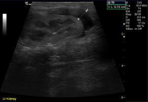
The left and right kidneys were severely enlarged with mild hydronephrosis in the right kidney. Both kidneys had hyperechoic cortices with large hypoechoic caps surrounding and invading the renal cortices (left kidney cap-7.4 mm in thickness, right kidney cap-13.6 mm in thickness).
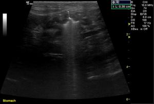
Stomach: mucosal layer is mild/moderately thickened with overall wall thickness measuring 3.8 mm
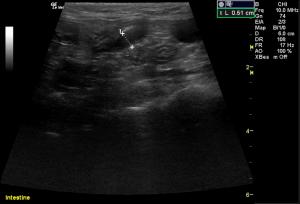
Intestine: there is focal mucosal layer thickening measuring up to 5.1 mm while the rest of the intestinal loops are of normal thickness.
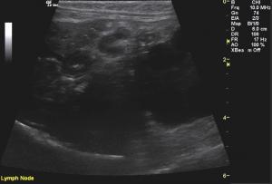
Lymph Nodes: Multiple mesenteric lymph nodes were severely enlarged throughout the abdomen. The epigastric LN enlargement is shown in this image in the near field.

History

Lymphoma Does What Lymphoma Does.

Anorexia, weight loss, “not doin’ right” are the usual signs in cats that make us suspect lymphoma or similar neoplasia but have us hope for pancreatitis, biliary disease, obstructive urolithiasis, orthopedic disease and other treatable disease with similar vague signs. However, when the uncontrolled cell growth of lymphoma strikes and causes every affected organ to “burst at the seams,” this is what the organs under the probe present like which we then sample accordingly. Dr, Marty Henderson of www.sonovet.us teams up with the amazing Claudia Barton DACVIM (Oncology) for the lymphoma diagnosis in this cat for the January, 2012 SonoPath Case of the month.

Sonogram (Liver): Lizzie

History: A 11-year-old female neutered DSH was presented with a history of anorexia and weight loss. The only abnormality on urinalysis is an inappropriate urine specific gravity ( 1.016). Azotemia is present on serum biochemistry.

Clinical Differential Diagnosis

(Lobetti DECVIM http://www.sonopath.com/specialists_lobetti.asp): Anorexia/weight loss – renal/hepatic/cardiac/pancreatitic disease, pyothorax, neoplasia, hyperthyroidism Renal disease – chronic kidney disease, neoplasia, pyelonephritis, hydronephrosis, renoliths, ureteroliths

Image Interpretation

(Henderson DVM, Lindquist DABVP)

Sonographic Differential Diagnosis

(Henderson DVM): Kidney Mass - the findings are severe -DDx: primary renal carcinoma, primary renal TCC, renal lymphosarcoma - may appear as diffuse disease or focal mass. Less likely: renal malignant histiocytosis or mastocytosis, malignant osteosarcoma, hemangiosarcoma . 2) Liver - the findings are moderately-severe - DDx: a) Hepatic lipidosis / Cholangiohepatitis b) Infiltrative neoplasia (lymphoma - probable in this case) c) Chronic vs. Acute hepatitis or cholangiohepatitis (bacterial vs. sterile vs. toxin) 3) Lymph nodes - the findings are moderate - DDx: infiltrative neoplasia is likely vs. reaction Due to the involvement of the liver, gastrointestinal tract, and multiple enlarged lymph nodes, lymphosarcoma is a highly likely.

Sampling

The right kidney and the liver were aspirated and slides are submitted for cytology review.

Outcome

No further outcome.

Comments

(Barton DACVIM): This cat clearly has disseminated lymphoma in all areas sampled, and one would presume that the other organs that were abnormal on the sonogram are also affected. Palliative prednisone or a chemotherapy protocol would be the treatment for this tumor, but the prognosis is very guarded to poor given the renal involvement. Many renal lymphomas in the cat spread fairly rapidly to the CNS, and thus lomustine/prednisolone might be a reasonable chemotherapy choice. Even with a 'good' response to chemotherapy, one would expect that remission duration would be 6 months or less, given the propensity of large-cell lymphoma in the cat to become chemotherapy drug resistant. The presence of numerous eosinophils among the neoplastic lymphocytes is interesting; it is likely that the lymphoid tumor cells are synthesizing interleukin 5. IL-5 is a key player in the coordination and orchestration of eosinophil-based inflammatory processes. There are occasional lymphomas in the cat in which the tumor cells apparently produce this cytokine, since the tumor tissue contains a very large number of eosinophils.

Videos

The liver was moderately enlarged, rounded in shape having mildly hypoechoic echogenicity with multifocal hypoechoic and cystic nodules throughout. The gallbladder was normal in size, shape, and clean.
The left and right kidneys were severely enlarged with mild hydronephrosis in the right kidney. Both kidneys had hyperechoic cortices with large hypoechoic caps surrounding and invading the renal cortices (left kidney cap-7.4 mm in thickness, right kidney cap-13.6 mm in thickness).
Stomach: mucosal layer is mild/moderately thickened with overall wall thickness measuring 3.8 mm
Colon: the colonic wall appeared thickened at the ileocecolcolic junction