Contact Us or call 800-838-4268 for more information
Skip to main content

GI Foreign Body

Patient Information

Age
8 Years
Species
Canine

Images

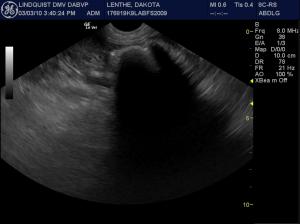
Case 1: A progressively shadowing, partially translucent luminal structure is present in the pyloric outflow.
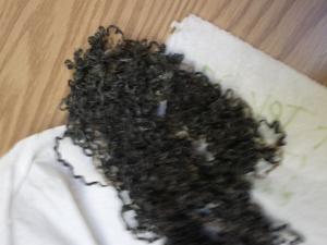
Case 1: Carpet foreign body was removed at gastrotomy. Image courtesy Doug Casey DVM, DABVP, Cert/Pres. IVUSS. English bay Imaging, Vancouver, BC, Canada. http://www.sonopath.com/specialists_casey.asp
Case 3: The mid descending duodenum reveals a distinctly shadowing and partially absorptive intestinal foreign body. Only a minor amount of luminal stasis is present suggestive for foreign material that will absorb fluid. Hyperechoic ill-defined fat is noted associated with the intestinal serosa suggestive for transmural inflammation in the affected portion of intestine.
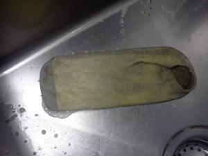
Case 3: A men’s running sock was retrieved at enterotomy.
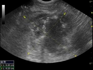
Case 4: The caudal aspect of the descending duodenum reveals a stiff, linear, echogenic luminal structure that penetrates the duodenal wall at the caudal flexure. The penetrating structure also creates a fistula into an abscess in the adjacent body wall measuring 5 x 6 cm.
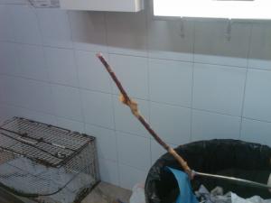
Case 4: A 10-inch stick was removed from the duodenum at surgery that was embedded in the caudal abdominal wall. The abscess was drained and lavaged. The patient recovered uneventfully. I still have no idea how that stick got past the pylorus!!!:)

History

Guess The GI Foreign Body Part 2

This month we provide more GI foreign bodies for your sonographic entertainment.

Guess the gastrointestinal foreign body!...Revisited…

Similar to last month, I give you the sonogram and brief history and you guess the foreign body that is present. Don’t scroll down until you think you know what the structure is.

Case 1: An 8-yr old Jack Russel terrier presented for persistent vomiting.

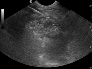
Case 2: A middle-aged FS Rottweiler presented for persistent vomiting after dietary indiscretion at a Sunday BBQ. Medical therapy was able to stop the vomiting but anorexia persisted.

Case 3: A 2-year-old Labrador retriever presents for vomiting and anorexia that is non responsive to medical therapy. Gastric and duodenal dilation with hyperperistalsis are present

Case 4: A 3 1/2-month old stray dog was brought to the veterinarian by a good Samaritan couple that found him by a garbage bin shocky, lethargic, and anorexic.

Comments

More information regarding the diagnosis of GI obstructions may be found in the resources tab on SonoPath.com where much of our research and articles are available. In particular see our abstract and PPT from ECVIM 2009 "Sonographic Criteria for the Diagnosis of Gastrointestinal Obstruction in 39 Dogs and Cats." Tons more GI obstruction and pseudo-obstruction cases are present in our searchable case archive for members of the SonoPath community. If you are not a member consider registering in our community, search out cases, post your video and case info to the community, and join the curve the SonoPath community lives for. Some of the images presented are part of the 400 images of sonographic pathology contained in the Atlas of Veterinary Sonographic Pathology-Small Animals and Exotics offered in the products page here at SonoPath.com.

Videos

Case 1: A progressively shadowing, partially translucent luminal structure is present in the pyloric outflow.
Case 2
Case 3: The mid descending duodenum reveals a distinctly shadowing and partially absorptive intestinal foreign body. Only a minor amount of luminal stasis is present suggestive for foreign material that will absorb fluid. Hyperechoic ill-defined fat is noted associated with the intestinal serosa suggestive for transmural inflammation in the affected portion of intestine.
Case 4: The caudal aspect of the descending duodenum reveals a stiff, linear, echogenic luminal structure that penetrates the duodenal wall at the caudal flexure. The penetrating structure also creates a fistula into an abscess in the adjacent body wall measuring 5 x 6 cm.