Contact Us or call 800-838-4268 for more information
Skip to main content

High Grade Lymphoma In A 12-Year-Old MN Labrador Retriever Mix: Our Case Of the Month September 2016

Patient Information

Age
12 Years
Gender
Male, Neutered
Species
Canine

Keywords

Exam Findings

Images

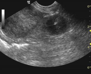
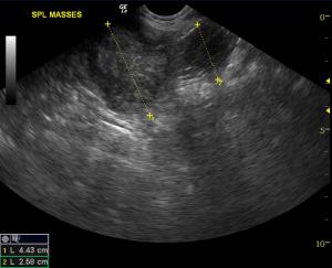
Splenic masses noted with parenchymal expansion and echogenic ill-defined pericapsular fat suggestive for an aggressive expansive process.
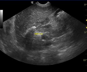
Hepatic lymph nodes enlarged but maintained the ovoid shape and normal length to width ratios. Reactive lymphadenopathy or infiltrative neoplasia are possible.
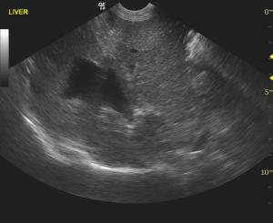
Liver: The Gb and CBD are normal. Parenchyma is mildly swollen with uniform parenchyma. The presentation is non specific consistent with benign hepatopathy or early infiltrative neoplasia given the splenic pathology and tendency for splenic spread of disease to the liver through the splenoportal system.
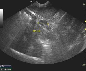
Splenic lymph node enlargement which is a concern for early pathological escape from the spleen.
US-guided FNA of the larger splenic mass.
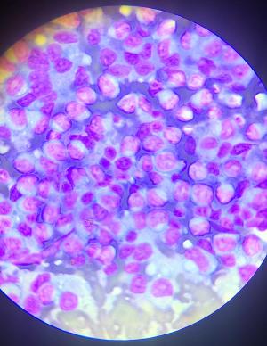
Telecytology sample demonstrating lymphoma cells obtained from the splenic FNA.

Consider a needle and a probe with telecytology to stage the patient for surgery vs medical therapy. This results in Enhancing Diagnostic Efficiency™ at its finest. This month's patient presented with splenic masses which could be a number of things (hematoma, hemangiosarcoma, round cell neoplasia, necrosis...) some of which are surgical and others that are not. With a needle, a probe, and SonoPath SDEP telecytology protocol within hours we knew this was a non surgical oncology case involving multiple organs with the ability to start chemotherapy within hours of presentation to the clinic. Ultrasound performed by Eric Lindquist DMV, DABVP, case managed by Dr. Alan Pomerantz and the staff of Franklin Lakes Animal hospital, telecytology read by Lawrence McGill DVM, PhD, DACVP.

History

A 12-year-old MN Labrador Retriever mix was presented with chronic diarrhea +/- syncopal episodes.Low grade anemia was found on CBC with unremarkable blood chemistry findings.

Image Interpretation

Spleen revealed multiple masses, the largest of which measured 4.43 and 2.58 cm with other hypoechoic nodules. The masses were moderately complex ,most consistent with sarcoma or similar. Other splenic nodules were noted measuring 2.8 cm. Splenic lymph node was enlarged at 1.6 cm. Liver in this patient was mildly enlarged, slightly coarse. Hepatic lymph nodes were slightly enlarged at 2 x 1 cm maintaining length to width ratios. Gallbladder and common bile duct were normal. Heart: Rapid view of the heart revealed no right auricular masses.

Sampling

FNA was sampled from both the spleen and liver.

DX

Splenic masses. Suspect round cell neoplasia or similar. Mildly heterogenous liver and hepatic lymphadenopathy. Regional lymphadenopathy medial to the spleen and in the portal hilus.

Outcome

3-view chest radiographs were recommended along with an echocardiogram. Given the syncopal episodes, serial blood pressures to assess for systemic hypertension were recommended. FNA of both the spleen and liver were performed without complication. Cytology interpretation: High grade lymphoma in liver and spleen.

Comments

Within 6 hours of the patient presenting for syncope and sonogram the definitive diagnosis was made to enable potential CHOP, Wisconsin or similar protocols to be implemented before further lymphoma spread could occur. The patient is currently (As of 8/27/16) undergoing chemotherapy and doing well so far.

For more information about SonoPath's telecytology service packages click here: TELECYTOLOGY

Videos

US-guided fna splenic mass. Woodpecker tecnhique.
US-guided fna liver screening for early infiltrative disease vs benign hepatopathy.
Spleen telecytology video demonstrative lymphoma cells.Excellent images of large numbers of rounded cells are submitted. The cells are large round lymphoid cells with nuclei that are mainly 2-4 times the size of RBCs. They have large nucleoli. The cytoplasm is dark and eccentric. There are very few mature lymphoid cells or neutrophils. This cellular collection is consistent throughout the images.
Liver telecytology video demonstrating lymphoma cells mixed within the hepatocellular population. The images are excellent collections of consistent cells that have the same pattern of those in the spleen. These cells are atypical lymphoid cells and blasts.