Contact Us or call 800-838-4268 for more information
Skip to main content

Histiocytic sarcoma in a 10-year-old FS English Bulldog: Our Case Of the Month September 2015

Patient Information

Age
10 Years
Gender
Female, Spayed
Species
Canine

Keywords

Clinical Signs

Special Testing

Images

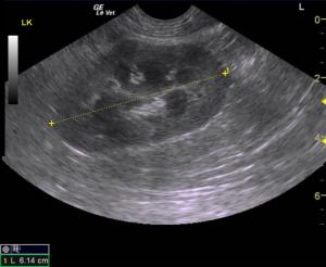
Left kidney with pericapsular fluid accumulation suggestive for an acute insult. Nephritis, leptospirosis, thromboembolic disease and neoplasia can all present in this manner.
Irregular right kidney. Corticomedulalary detail loss and multifocal capsular expansion and early escape noted with echogenic pericapsular fat suggestive for an expansive neoplastic or aggressive inflammatory process.
FNA of right kidney with 25 gauge needle.
An image of tumor cells from the right kidney demonstrating variable sizes and shapes of nuclei with chromatin clumping. Nucleoli are present. The cytoplasm of the tumor cells is varied (anisocytosis). Some of the cytoplasm is staining quite dark. This is an excellent image with good collections of cells.
A good clear image of cells from the right kidney with variable sizes and shapes. Scattered neutrophils and lymphocyte are present in the collection. One of the cells is binucleate. These are atypical tumor cells with clumped chromatin and prominent nucleoli.
This image from the FNA of the right kidney, demonstrates a variation in nuclear size and shape. (Anisokaryosis) the nuclei demonstrate clumped chromatin. The cytoplasm is difficult to identify in some cells. There is aggregation and clumping of these cells.

A small irregularity of a kidney deserves a needle. Kelly Vazquez, CVT of SonoPath Mobile New Jersey, scans this "ADR" English Bulldog with evidence of UTI with a Lindquist virtual read. Even though UTI is evident in the history, the needle in the renal cortex says something completely different through telecytology. Those who wield a needle well win the diagnostic efficiency challenge and this case proves that concept once again. Case management by Dr. Chris Hallihan of Animal Care Center Flanders, in Flanders, NJ. Cytology interpretation by Dr. Larry McGill of SonoPath telecytology services.

History

A 10-year-old FS English Bulldog was presented due to diarrhea, decreased appetite, and vomiting 3-4 times in a day. An interstitial lung pattern was seen on radiographs. Upon physical examination the patient was QAR with a painful abdomen upon palpation. A CBC found WBC 18.8, neutrophils 15416, monocytes 1692, and 0 eosinophils. A blood chemistry found amylase 1678, TP 7.7, globulins 4.4, Creatinine 2.0, and anaplasma positive. A urine culture was negative and a urinalysis showed a specific gravity of 1.007, protein +2, Blood +3, WBC 11-20, and RBC 21-50.

Image Interpretation

Normal echocardiogram for this breed. Abdominal sonogram: Irregular kidneys with pericapsular inflammatory pattern. Corticomedullary detail loss and pericapsular ill-defined echogenic fat noted more prominently in the right kidney with subtle architectural disruption. pericapsular fluid noted around the left kidney suggestive for acute insult. Trace amount of pleural effusion was noted through the diaphragm. Thickened gastric wall with artifact.

Sonographic Differential Diagnosis

Nephritis gastritis vs emerging nephric neoplasia of round cell origin. Recommend US-guided FNA to further define mixed inflammation of the renal cortices vs round cell infiltrative disease that would yield a monopopulation.

Sampling

US-guided 25 g FNA revealed histocytic sarcoma of the right kidney.

Outcome

Cause of the effusion could be paraneoplastic or owing to systemic inflammatory response. Guarded prognosis depending on underlying cytology. Aggressive treatment for nephritis is recommended. Gastrointestinal protectants, broad spectrum antibiotics, sedation and 25-gauge FNA of the right kidney would be recommended. U/S guided FNA of the right kidney was performed without complication.

Comments

Pathology results from U/S guided FNA of the right kidney found: Neoplastic cells suggesting round cell tumor or extremely pleomorphic carcinoma. Telecytology interpretation was performed by Dr. Larry McGill, Ph.D Diplomate, ACVP Medical Director (Emeritus) and Veterinary Pathologist. 

Videos

Left kidney.
Irregular right kidney.
FNA of right kidney.
Right kidney post FNA.