Contact Us or call 800-838-4268 for more information
Skip to main content

Intestinal Mast Cell Tumor in an 11-year-old MN DSH Cat: Our Case of The Month May 2016:

Patient Information

Age
11 Years
Gender
Male, Neutered
Species
Feline

Keywords

Clinical Signs

Images

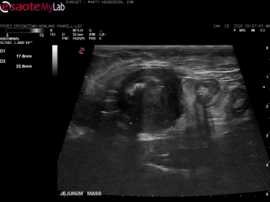
Jejunal mass.
Normal layering and thickness of intestine.

Palpable mass in a cat?… what can it be? It’s not always intestinal lymphoma as other round cells can be involved such as mast cell disease… or even non neoplastic pathology such as complicated IBD, spontaneous necrosis, bowel infarction, or even FIP. Have a probe & a needle & Marty Henderson DVM of http://sonovet.us/ will get the answer. Take a look at another variant of the ropey bowel cat pathology and why a needle and a probe are always in need in this SonoPath case of the month.

History

This 11-year-old MN DSH cat vomited all of his food 3 days ago. Owner noticed grass-eating and some vomiting of grass last week. Normal activity and appetite. Currently on Cerenia and SQ fluids. R/O FB vs pancreatitis vs other.

Image Interpretation

Most loops of the intestines are WNL - normal bowel layering, wall thicknesses and motility. No mucosal stippling or disproportionate layering appreciated. There is a large hypoechoic heterogeneous mass measuring ~2cm involving the mid-jejunum in the lower right quadrant. The mass is well-circumscribed but the jejunum leading up to the mass has abnormal thickening of the muscularis layer and tapers to normal. The layering in the mass is obliterated and the mass is mildly erupting into the bowel lumen. Hyperechoic ill-defined reactive mesentery is associated with the mass suggestive for an aggressive transumural pathology with associated inflammation/peritonitis. Multiple mesenteric lymph nodes are moderately-severely enlarged and abnormally shaped having homogenous hypoechoic echogenicity.

Sonographic Differential Diagnosis

1. Intestinal Mass - the findings are moderately-severe - DDx: adenocarcinoma vs. infiltrative neoplasia (lymphosarcoma vs. mast cell tumor) vs. feline gastrointestinal eosinophilic sclerosing fibroplasia (FGESF) vs. leiomyosarcoma vs. leiomyoma, dry form FIP possible but less likely. 2. Lymph nodes - the findings are suggestive of metastasis from the intestinal mass - DDx: infiltrative neoplasia (lymphoma vs. mast cell vs. other) vs. IBD vs. infection vs. reaction vs. metastatic neoplasia.

Sampling

US-guided biopsy of mass was performed.

DX

Intestinal mast cell tumor. *

Outcome

Biopsies recommended to accurately define the type of disease present. Consider surgical resection with histopathology and appropriate chemotherapy. Prognosis is very guarded. Pathology findings: *Transmural mast cell tumor with invasion of adjoining mesentery. Margins: Appears completely excised with wide margins. Vascular invasion: Not detected. Comments: The submitted jejunum contained a transmural mast cell tumor that appeared to have been completely excised but could have some potential for metastasis to other sites including the mesenteric lymph nodes and the liver. Intestinal mast cell tumors in the cat may be solitary yet are often accompanied by local lymph node and occasionally hepatic and splenic involvement. Close monitoring for any signs of metastasis is advised.

Comments

Scan by Marty Henderson DVM of SonoVet mobile ultrasound, a special thanks to him for providing a fantastic set of images.

Videos

Jejunal mass.
Jejunal mass with loss of mural detail and surrounding reactive mesentery.
Enlarged, irregular, hypoechoic mesenteric lymph nodes. The normal length to width ratio is distorted.