Contact Us or call 800-838-4268 for more information
Skip to main content

Invasive Adrenal Mass In An 8-Year-Old MN Pit Bull Terrier: Our Case Of the Month December 2022

Patient Information

Age
8 Years
Gender
Male, Neutered
Species
Canine

Keywords

History

Clinical Signs

Urinalysis

Images

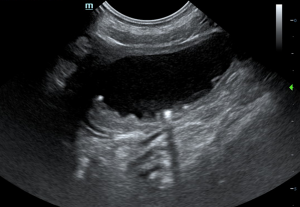
Dependent sand along the urinary bladder wall.
Left adrenal gland with part of mass above it.
Suspect pheochromocytoma (left).
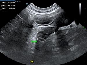
Normal right adrenal gland.

We don't call her the "Adrenal Queen" for nothing. Diane McFadden, BS, RVT, SDEP® Certified Clinical Sonographer, Director of Mobile Operations and Education for SonoPath, is from our team of highly skilled sononographers that scan for Sonopath Mobile Veterinary Ultrasound (northern New Jersey). Diane's beautifully diagnostic images of this suspect pheochromocytoma were interpreted by Eric Lindquist, DMV, DABVP, Cert. IVUSS. and demonstrate how the adrenal mass is invading into the CVC. Dr Silas Ashmore of All Creatures Great and Small Animal Hospital and Urgent Care managed this case.

History

An 8-year-old MN Pit Bull terrier was presented for chronic hematuria over the past year. The urine is noticeably more red following exercise. No current meds. Urinalysis revealed only increased red blood cells.

Image Interpretation

The urinary bladder, trigone, and pelvic urethra presented normal thicknesses and normal tone. The ureters were not visible which is normal. A minor amount of suspended debris was noted along with a minor amount of dependent sand. A grouping of which measured 0.6 cm and was non-obstructive.

The left adrenal gland revealed a portion of the left adrenal gland that expanded into a vascular mass with phrenic vein invasion. The mass extended into the vena cava approximately 3-4 cm. The pattern is strongly suggestive of pheochromocytoma. The left adrenal gland was imaged from the left and right approaches. The left adrenal gland measured 3.35 x 0.91 cm at the caudal pole and 0.86 cm at the cranial pole. The right adrenal gland was normal and measured 1.06 x 0.62 cm at the caudal pole and 2.54 cm in length.

DX

Left adrenal mass, invasive through the vena cava. Suspect pheochromocytoma.

Outcome

CT evaluation would be warranted for potential surgical planning or direct surgical intervention if the surgeon is prepared for vascular invasion approximately 3.0 cm cranially into the vena cava through the left phrenic vein. There was no organ metastasis. Urine catecholamine is warranted as well as serial blood pressure measurements and chest radiographs.

Videos

note the phrenic vein invasion into the mass
It's important to image left adrenal abnormalities from the right side to fully assess. From the right adrenal, move the probe slightly caudally and angle.
Even if the left adrenal appears normal, always image the CVC from the right side for any "surprises"