Clinical Differential Diagnosis
(Remo Lobetti PhD, DECVIM): Neoplasia, pyometra, chronic inflammatory disease, gastritis, pancreatitis, bowel torsion, abdominal or thoracic hemorrhage.
Image Interpretation
(Lindquist DMV, DABVP): The uterus was severely dilated with echogenic fluid with thickened, irregular and polypoid changes (Videos 1 & 2). This is consistent with chronic pyometra. The dilated uterus occupied most of the abdomen. The ovaries were not visualized. The sonographer must ensure that these dilated tubular structures are not intestine or severely dilated ureters that can also, in extreme circumstances, appear similarly during obstructive pathology in their respective organ systems. These dilated "tubes" connected caudally with the cervix and the uterine body was appropriately positioned between the bladder ventrally and the colon dorsally in the pelvic inlet in order to ensure that pyometra was the correct diagnosis.
Sonographic Differential Diagnosis
(Lindquist DMV, DABVP): Pyometra with concurrent peritonitis or hemorrhage
Sampling
Immediate exploratory surgery was performed with a rapid ovariohysterectomy.
Outcome
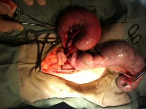
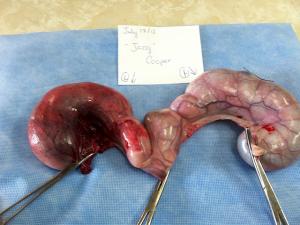
Torsion of the left horn was found with hemoabdomen owing to resultant hemorrhage (images 5 & 6). The patient recovered uneventfully.


Comments