Contact Us or call 800-838-4268 for more information
Skip to main content

Left Adrenal Mass in a 16yr old, MN, Havanese

Patient Information

Age
16 Years
Gender
Male, Neutered
Species
Canine

Keywords

Clinical Signs

Images

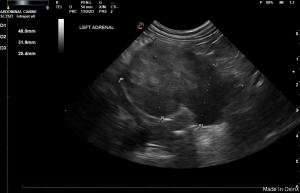
The tumor of the left adrenal gland can be seen here measuring 48mm x 24mm.
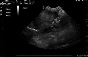
The normal right adrenal gland can be seen here as a comparison.
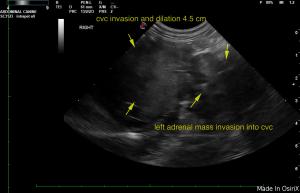
The adrenal mass invasion into the CVC.

History

A 16-year-old MN Havenese with a history of an adrenal tumor was presented for evaluation of chronic intermittent vomiting for the past 4 weeks. Current therapy was Lysodren, famotidine, carafate, enalapril, amlodipine, and tramadol 50mg ¼ BID.

Clinical Differential Diagnosis

Drug reaction Addison’s disease Organ failure/disease – liver, kidney, pancreas GI tract – neoplasia, inflammatory bowel disease, foreign body, parasitic enteritis Neoplasia

Image Interpretation

The urinary bladder, trigone, and pelvic urethra presented normal thicknesses and normal tone. The ureters were not visible which is normal. Bladder stones were noted. No evidence of inflammatory or neoplastic changes was noted.  Ureteral papillae were normal.

The left kidney measured 4.4 cm with multiple calculi, cyst , moderate degenerative changes and infarct. The right kidney was uniform and measured 4.47 cm.

The left adrenal gland mass now measured 4.8 x 3.2 cm and was irregular in contour.  The tumor now invaded the vena cava to an extent of 4.3 cm of dilation of the vena cava.  The right adrenal was uniform and measured 1.72 x 0.57 cm cranial pole and 0.63 cm at the caudal pole.

The base and limbs of the pancreas were observed to be largely isoechoic to surrounding omental fat. Some parenchymal remodeling, however, with mild deviation from curvilinear normalcy was observed. Pancreatic duct and capsular irregularities were present consistent with age related changes. If pain upon imaging (+ Murphy sign) was present or if the patient is focally painful in subxiphoid palpation then low-grade smoldering chronic pancreatitis should be suspected.

Sonographic Differential Diagnosis

Progressive left adrenal mass with invasion into the vena cava. The mass was significantly vascular. Age related pancreatic changes. Bladder stones. Left renal stones, cyst, and infarct.

DX

Left adrenal mass with invasion into the vena cava

Outcome

Palliative therapy is recommended. Some inspissated blood was noted in the vena cava in this patient. There is a high potential for clot/thromboembolic disease. Serial blood pressure measurements are recommended. Clinical support is recommended regarding the clinical signs.

Videos

The tumor of the left adrenal gland is clearly seen here.