Contact Us or call 800-838-4268 for more information
Skip to main content

Linear foreign body Obstruction in a 3-year-old FS Pot Bellied pig: Our November Case Of the Month 2015

Patient Information

Age
3 Years
Gender
Female, Spayed
Species
Exotic

Keywords

Blood Chemistry

Exam Findings

Images

"Taylor Ham" for Thanksgiving? Not if Dr. Paul Sedlacek and his scanning wondertechs Jennifer Nolan & Pam Tosto with Virtual Lindquist on interpretation have anything to do with it. :) "Taylor Ham,” (Yes, that’s her name!) the 3-year-old domesticated Pot Bellied Pig presented to Animal Clinic of Morris Plains with GI signs and, well... a history of "dietary indiscretion.”  It's a pig so how do we define "dietary indiscretion??" Anyway see what we found with the SonoPath trained scanning technicians at ACMP in Morris Plains, New Jersey, USA.

History

The patient was presented for evaluation due to severe vomiting. The vomiting stopped but the patient was now anorexic. The patient was quiet with a thin body condition. CBC found a low WBC of 3.7. Blood chemistry found albumin 3.7, total protein 7.0, ALKP 43, BUN 43, and cholesterol 79.

Image Interpretation

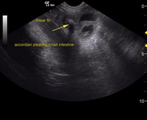
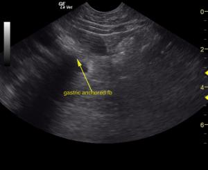
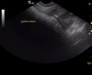
The stomach revealed shadowing foreign material. The gastrointestinal tract revealed linear foreign body with accordion pleating. Dilated bowel was noted. Severe gastric stasis was noted. Minor, regional hyperechoic fat was noted along with slight free fluid accumulation. Further material was lodged in the distal small intestine connecting to the material within the pylorus. The remainder of the abdomen was unremarkable. Slight free fluid was noted consistent with emerging peritonitis.

DX

Linear foreign body with accordion pleating and emerging peritonitis.

Outcome

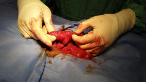
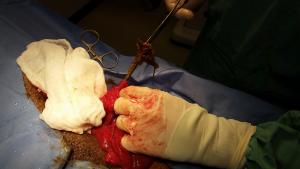
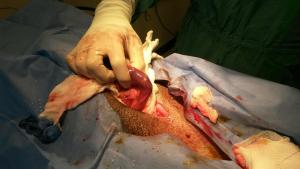
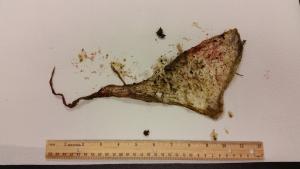
The patient was recommended for immediate exploratory surgery. Surgery was performed with two enterotomies, jejunum and duodenum. Plastic wrap was removed from both locations. The plastic had twisted into a linear shape. The duodenal foreign body extended into the stomach.   

Videos

Echogenic gastric material demonstrates progressive shadowing followed by small intestinal luminal dilation tethered by an echogenic linear foreign body. Accordion pleating is seen as well as empty small bowel in the 3rd video with the linear material traversing the pleated bowel at a 90 degree angle. Regional echogenic fat suggest emerging peritonitis. Gastrointestinal fabric type linear foreign body and emerging peritonitis.