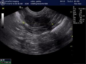
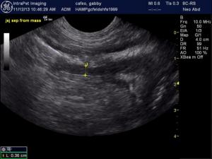
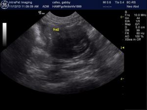
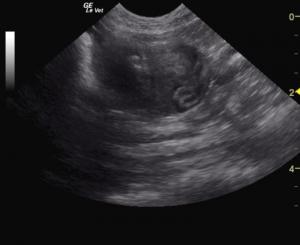
Sneaky mast cell disease in cats. Looks like lymphoma, acts like lymphoma but you don't need a lot of MCT for significant clinical signs owing to degranulation especially in cats. Look at rock star veterinary sonographer Andi Parkinson, RDMS of Intrapet Imaging, Baltimore, MD, USA guide her client in an US-guided FNA into this feline intestinal mass avoiding the lumen and nailing the Dx. Just push the mass to the skin, minimizing the real estate, and stick… its like placing the cue-ball close to the 8-ball when playing billiards. :) See our video on how to perform an FNA here.
Clinical Differential Diagnosis
Neoplasia, abscess, foreign body, FIP.
Image Interpretation
A focal intestinal mass is seen and progressive infiltrative pattern with loss of mural detail. Partial obstructive pattern is noted with trapped echogenic luminal ingesta prior to the mass.
Sonographic Differential Diagnosis
Intestinal mass: Lymphoma, MCT, leiomyosarcoma, complicated inflammatory disease, FIP.
Sampling
US-guided FNA: Mast cell neoplasia.
DX
Intestinal mast cell neoplasia.
Outcome
The patient was referred to an oncologist for evaluation. No further medical intervention was sought and at the time of the publication of this case the patient was still stable.


