Contact Us or call 800-838-4268 for more information
Skip to main content

Restrictive Cardiomyopathy

Patient Information

Age
9 Years
Gender
Female, Spayed
Species
Feline

Images

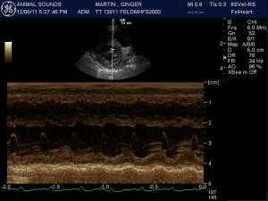
Left atrial enlargement is confirmed in the La/Ao mmode of 1.9. EPSS is mildly excessive at 0.4 cm. The left ventricular cavity is mildly dilated in diastole (1.9 cm) and systole (1.4 cm) with a depressed fractional shortening (26 %) consistent with myocardial failure. The left ventricular walls are normal (0.4 cm septal wall and 0.4 cm free wall). The heart rate is measured at 184 to 206 bpm and a regular sinus rhythm is seen on an ECG run during the exam.
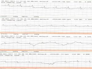
The ECG shows a regular sinus rhythm with a mildly elevated rate of 200 bpm. There are no arrhythmias on this recording. The QRS complexes are towards the upper limit of normal and the rest of the P-QRS-T complex appears normal.
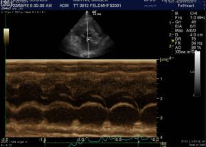
(Follow up image) La/Ao m-mode is normal with a ratio of 1.3.
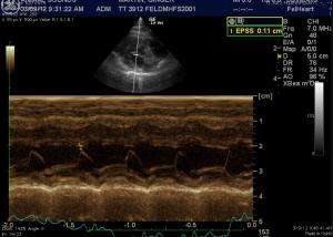
(Follow up image) LV m-mode EPSS has also improved from 0.4 cm to 0.11 cm which is normal for a cat.

History

The Restrictive Cardiomyopathy Cat, a Saddle Thrombus, & Today’s Cutting Edge Effective Pharmacy.

Yes the kitchen sink of medications were necessary to pull this cardiomyopathy cat through a saddle thrombus. Plavix, pimobendan, ace-inhibitor, atenolol, lasix, heat support and whatever else came to mind….A fortunate 21st century cat and excellent care prevailed thanks to a trio of local intensive care, mobile sonography, and remote cardiology consultation. You have to love where we are in the “Flat” veterinary world of 2012.

Sonogram (Cardiac): Ginger Martin

History: The patient is a feline DMH, SF, 9 year old. The patient has a history of hyperthyroidism, treated with I 131, sudden onset posterior paresis. The physical exam revealed poor femoral pulse quality, knuckling of hind limbs, gallop rhythm, and hypothermia (97.2 F). The patient was able to move the rear paws but could not stand. Bloodwork revealed slight hyperglycemia, moderate anemia, polychromasia and anysicytosis. Thrombocytopenia with clumping was also noted.

Clinical Differential Diagnosis

(Remo Lobetti PhD, DECVIM): Saddle thrombosis secondary to cardiac disease - dilated cardiomyopathy, hypertrophic cardiomyopathy, restrictive cardiomyopathy Spinal - trauma, disc, neoplasia Myopathy - trauma, infectious

Image Interpretation

(J. Betkowski DVM, DACVIM Cardiology):

Sonographic Differential Diagnosis

(J. Betkowski DVM, DACVIM Cardiology): The echocardiogram is consistent with a restrictive cardiomyopathy with moderate left atrial enlargement, an episode of early hear failure and a thromboembolic event. This condition is likely not related to the hyperthyroidism diagnosed earlier. It appears relatively advanced at this time and puts the cat at increased risk for a repeat thromboembolic event. A persistently elevated heart rate can also contribute to signs of decompensation.

Sampling

(J. Betkowski DVM, DACVIM Cardiology): Heparin can be considered at 200 U/kg IV once followed by 100 U/kg SQ QID while the cat is hospitalized. Other supportive care such as pain medications can be considered as clinically warranted. Low dose Lasix such as 6.25 to 7.5 mg IV q 12 to 24 hours can be considered, particularly if the respiratory rate were elevated. This can be continued as a maintenance therapy, perhaps at 6.25 mg PO SID or whatever is the lowest dose that will control the clinical signs. Pimobendan is also recommended at 0.625 to 1.25 mg PO BID. Once the cat has improved, Benazepril can be added at 1.25 mg PO SID and increased to 2.5 mg PO SID after a week if it is well tolerated. If the heart rate were persistently elevated over 180 bpm, Atenolol would be recommended at 1/4 of a 25 mg tablet PO SID to BID to keep the heart rate in the normal range. Long-term anti-coagulation can be considered with aspirin or Ascriptin at 5 mg/kg or 5 mg total dose PO on a Monday, Wednesday, Friday schedule with Plavix at ¼ of a 75 mg tablet PO SID. A renal profile is recommended a week after the final adjustments to the medications have been made to make sure they are well tolerated. The condition can be followed by thoracic radiographs every 2 to 5 months to monitor for signs of progression or decompensation. The echocardiogram can be repeated in 6 months to monitor for any further changes to the cardiac structure.

Comments

Special thanks to Tomie Timon RDMS of Animal Sounds Mobile Veterinary Ultrasound in Eugene, Oregon, USA, Dr. Sheri Schlorman & staff at Creswell Veterinary Hospital, Creswell, Oregon, USA & Dr. Jean Betkowski DVM, DACVIM-Cardiology for their excellent management of this cardiac patient.

Videos

The left atrium is moderately enlarged in size. All valves viewed appear normal and the motion of the mitral valve. There is mild mitral regurgitation noted on color flow Doppler and no other abnormal flows are seen. The left ventricular papillary muscles appear normal. The right ventricle is prominent and the right atrium is normal to mildly enlarged in size. The main pulmonary artery appears prominent in comparison to the aorta.
Heart Based & Oblique 5-Chamber Views: Left atrial enlargement is confirmed in the heart-based view. There is no pleural effusion and a scant volume of pericardial effusion is present in the 7-10 O’clock position in the heart based and oblique 5-chamber view. There is no evidence of neoplasia on this exam.
The 5-chamber long axis, heart base, LV short axis mushroom view, and apical views demonstrate normalization of LA and LV volumes with improved ventricular kinesis with the current therapeutic protocol.