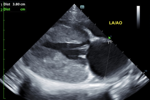
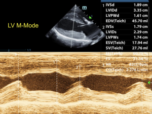
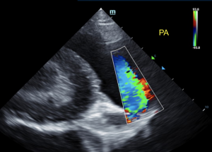
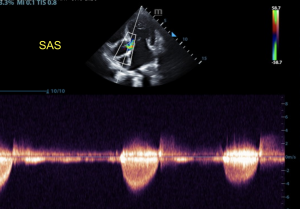
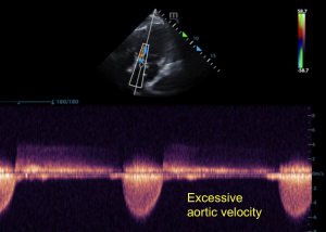
"Subaortic stenosis is one of the most common congenital heart defects in dogs, characterized by an obstruction of the left ventricular outflow tract, resulting in pressure overload on the left ventricle. It is caused by fibrous nodules, a ridge or ring of fibrous or fibromuscular tissue located just below the aortic valve, which increases resistance to left ventricular outflow. It most commonly occurs in large breed dogs such as Newfoundlands, Boxers, Rottweilers, Golden Retrievers, Dogue de Bordeaux, German Shepherds, German Shorthaired Pointers, Great Danes, Bouvier des Flandres, and Mastiffs, and appears to be genetic in origin. Signs of subaortic stenosis can be present at birth or develop during the first year of life. Depending on the degree of resistance to left ventricular outflow and the resulting increase in outflow velocity, it can be graded as mild, moderate, severe and very severe (extreme)." - Peter Modler. See what a heart with subaortic stenosis looks like in a not-so-little 9-month-old male Mastiff mix puppy. Diagnostic imaging provided by Jenna Walsh, CVT of Animal Sounds Northwest, image interpretation by Eric Lindquist, DMV, DABVP, Cert. IVUSS, concise description of subarotic stenosis by Peter Modler, DVM, Dipl.-Tzt., Specialist German Board of Cardiology
DX
Subaortic stenosis with concurrent mitral and tricuspid insufficiency - Secondary left ventricular hypertrophy. - Periodic arrhythmia.
Outcome
Atenolol therapy was recommended in this patient depending upon EKG results. Guarded to poor long-term prognosis. Blood pressure measurements were indicated if not already performed. Follow up with the patient's RDVM was unfortunately not available at the time of this study posting, we hope he is doing ok.

