Clinical Differential Diagnosis
(Lobetti BVSc, MMedVet, PhD, DECVIM):
Depression/anorexia - non-specific
Splenomegaly - neoplasia/torsion/infarction/primary splenic
infection/systemic infection/inflammation
Neutrophilia - infection/inflammation/neoplasia/immune-mediated disease
Image Interpretation
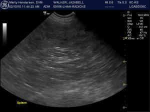
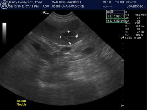
(Lindquist DMV, DABVP): Sonogram 1: Severely mottled echotexture with multifocal hypoechoic changes and several discrete complex hypoechoic nodules throughout. The overall splenic shape is irregular with capsular deviation suggestive for an aggressive pathological process. Sonogram 2: The largest discrete nodule is measured as a benchmark for future rechecks to assess any progression of the pathological process.
Sonogram 3: Video clip assessment of the spleen better defines the diffuse nodular changes and scalloping capsular contour suggestive of a relatively aggressive underlying pathology.
Sonogram 4: Further video clip assessment of an exemplary nodule in the spleen. Note the loss of architectural detail in that the curvilinear aspects of normal splenic parenchyma are completely lost owing to the underlying pathology. Sonogram 5: Color flow Doppler assessment of the splenic vasculature is adequately curvilinear yet without significant impingement from adjacent nodular changes. This is interesting in that usually, in my experience, neoplastic nodular changes are usually expansive within the architecture of the affected organ such as the capsule in this case. But this also usually includes deviation of vessels within the organ itself creating mini "mass effects" within the organ examined. This is not the case in this patient as the vessels maintain their normal straight contour within the spleen. Infiltrative disease (lymphoma, hemangiosarcoma, mast cell neoplasia....) is, of course, in the primary differential list given the parenchymal architectural deviation and scalloping splenic capsule. However, given the maintained linearity of the vasculature evidenced in color flow Doppler, an aggressive inflammatory or severe nodular hyperplasia pathology is to be considered as well.
Sonographic Differential Diagnosis
(Henderson DVM):
Splenomegaly Diffuse
a) Inflammation (Splenitis)
1. Suppurative
2. Necrotizing
3. Eosinophilic
4. Lymphoplasmacytic
5. Granulomatous
6. Pyogranulomatous
b) Hyperplasia
1. Infection
2. Immune-mediated disease
3. Congestion
c) Infiltration
1. Neoplasia
2. Extramedullary hematopoiesis
3. Amyloidosis
Sampling
US-guided fine needle aspiration (Henderson DVM).
Cytological Interpretation (Barton DVM, DACVIM-Oncology/Internal Medicine):
Spleen - Moderate to marked extramedullary hematopoiesis, with focal pyogranulomatous inflammation suggestive of microabscesses; no evidence of lymphoma or other neoplastic infiltrate.
Outcome
The patient underwent splenectomy and long-term antibiotic therapy and recovered uneventfully. No cultures were performed owing to cost concerns.


Comments