Contact Us or call 800-838-4268 for more information
Skip to main content

Severe Chronic Inflammatory Process In A 10-Year-Old MN DSH Cat: Our Case Of the Month Janaury 2022

Patient Information

Age
10 Years
Gender
Male, Neutered
Species
Feline

Keywords

Clinical Signs

Exam Findings

Images

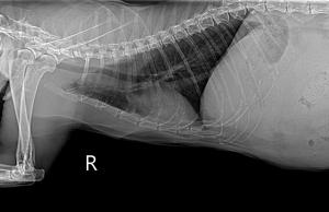
Right lateral thoracic radiograph.
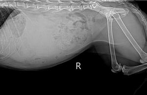
Right lateral abdominal radiograph.
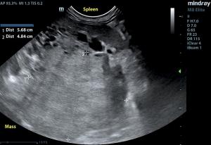
Splenic mass.
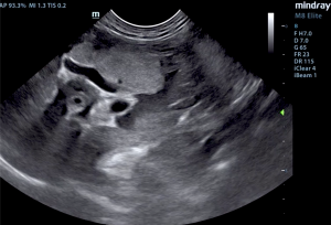
Mass associated with area of the pancreas.
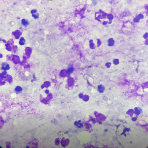
Cytology image from FNA shows neutrophils and degenerate cells in the mass collection from the patient. There are suggestions of bacteria in the background

Karen Ebersole, DVM, SDEP® certified clinical sonographer/instructor, and owner of Scanvet Mobile Veterinary Ultrasound (which provides services in central Maine, from Portland to Augusta), performed the ultrasound for this month's Case Of the Month. She hit the mark with her fine needle aspirates and processing of her diagnostic slides utilizing telecytology;  as we say here at SonoPath, "Nice shootin Tex!". Dr. Ebersole was able to provide the answers needed to her client in order to decide on the best treatment options for this patient. Intepretation of ultrasound images by Eric Lindquist, DMV, DABVP, Cert. IVUSS founder & CEO of SonoPath.com. Cytology read by L.D. McGill, DVM, Ph.D., DACVP.

History

A 10-year-old male neutered FIV (+) DSH cat was presented for a mass palpated in the cranial abdomen. Weight loss of >2lbs. but acting normally otherwise. Physical exam found the patient with a BCS of 4/5 and a large spherical mass palpable in cranial abdomen. Radiographs showed a mass effect caudal to stomach with the intestines displaced caudally.

Image Interpretation

Ultrasound: The spleen revealed a complex mixed echogenic cystic and hyperechoic parenchymal mass measuring approximately 7+ cm. The mass appeared fairly vascular and occupied much of the mid cranial abdomen. The mass appeared to be deriving from the caudate process of the liver; however, it occupied areas of the pancreas as well. The gastrointestinal tract was deviated by the mass. Free fluid noted in the abdomen, likely owing to mass hemorrhage.

Radiographs: Radiographs revealed undifferentiated cranial abdominal mass and ascites.

Cytology: Submitted are several videos and one image collected from the abdominal mass in the patient. The cellularity is rather uniform throughout all of the videos and the one image consists of necrotic cells. There is necrotic debris interspersed with prominent neutrophils and very few other inflammatory cells. Many of the neutrophils are demonstrating toxicity or degeneration. There are scattered bacteria in the background. In the more hemorrhagic collection, the cellularity is mixed but it is inflammatory. Neutrophils are prominent in those videos as well. The cellularity in all of these videos supports a rather severe chronic inflammatory process. There are excellent collections of cells that support an abscess. There are even scattered structures that suggest coccobacillus bacteria. There is no suggestion of malignant cellularity in the collection.


Sampling

Ultrasound guided FNA of the mass was performed without complication.

DX

Ultrasound: Undifferentiated mass in the region of the pancreas associated with the spleen and caudate liver, deviating the gastrointestinal tract. Cytology: Abdominal mass - Severe chronic suppurative inflammation consistent with an abscess and bacterial infection.

Outcome

The patient is currently being managed with antibiotics and is stable at this time.

Comments

Comments and recommendations from ultrasound: The mass does not appear resectable. It impinges upon the spleen and could be deriving from the spleen, yet would be an odd pattern for a splenic mass. The mass is likely adhesive to the spleen and not necessarily deriving from it. Exploratory surgery could be considered depending upon the results of the FNA. Chronic suppurative necrotic disease possible depending upon cytology results.

Comments and recommendations from cytology: The cellularity in all of these videos supports a rather severe chronic inflammatory process. There are excellent collections of cells that support an abscess. There are even scattered structures that suggest coccobacillus bacteria. There is no suggestion of malignant cellularity in the collection. Treatment with antibiotics and elimination of the mass may be beneficial for treatment in Tank. This easily could be derived from the pancreas but it could be from other sources as well. A guarded prognosis is warranted in these cases of rather severe inflammation in the abdomen of cats.  Culture may be beneficial if material is available.

Videos

Complex mixed echogenic cystic and hyperechoic parenchymal splenic mass.
Power Doppler of splenic mass.
Mass in the region of the pancreas.
Mass impinging on the liver.