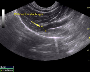
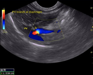
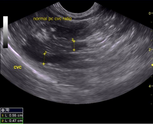
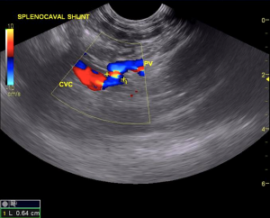
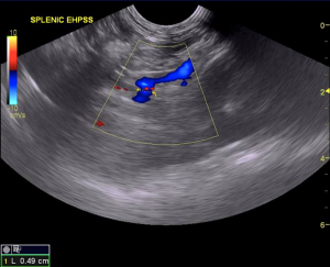
A smaller than average cat or dog, failing to thrive, with high post-prandial bile acids all points to a likely liver shunt. It is critical to be able to obtain solid images of the liver and the portal hilus to acheive the diagnosis. Dr. Eric Lindquist imaged this smaller than average feline patient and identified a definitive splenocaval shunt utilizing SDEP™ positions 13 and 14 along with Doppler evaluation. Diagnosis, corrective surgery, and the cat is doing well post-op and thriving. Special thanks to Dr. Kristen Casulli, Dr. Chris Hallihan, and the attentive staff at Animal Care Centers of Flanders for the management of this case.
Every sonographer can easily develop the skills to rule-in or rule-out both intrahepatic and extrahepatic shunts. The technique is built into the SDEP™ 17-point protocol that SonoPath teaches at all of our 3 day SDEP™ abdominal lecture and wet labs. For more information or to register for one of our 2019 labs click here: 2019 SonoPath SDEP™ Veterinary Ultrasound Training
DX
Splenocaval shunt with normal portal vein to vena cava ratio. Minor microhepatica.
Outcome
Recommend surgical intervention with ameroid constrictor therapy and concurrent liver biopsy +/- renal biopsy. The patient underwent shunt repair surgery with ameroid ring constrictor placement and did well post-operative. Follow up blood work found all values completely normalized. The patient continues to thrive at home and at his last wellness check up he weighed 11.6 pounds compared to his pre-shunt treatment weight of 6 pounds.

