Sonographic Differential Diagnosis
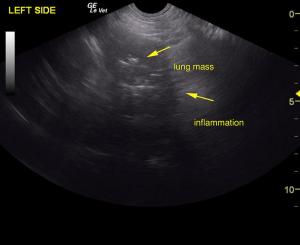
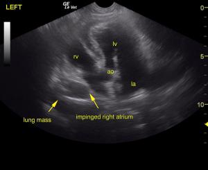
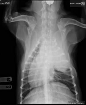
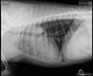
Left cranial thoracic mass. Echotexture likely lung origin, less likely thymoma or ectopic thyroid carcinoma.
Sampling
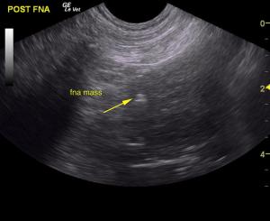
U/S-guided FNA of thoracic mass was performed without complication.
DX
Cytology is consistent with a pulmonary adenocarcinoma.
Outcome
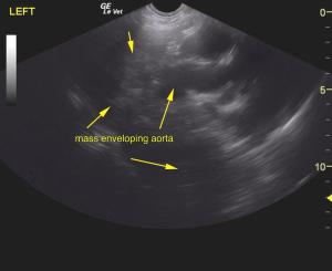
Given the rapport with the aorta and other regional vasculature, resection will likely be difficult. However, CT evaluation would be ideal in this case for potential surgical planning depending on cytology results. Lung carcinoma or possible thymoma are primary differentials. If lung origin, differentials include lung carcinoma, lung sarcoma and lung necrosis. This may be thymic in origin. Radiology review of the radiographs would be ideal with oncology consultation if neoplasia is confirmed on the surgical and/or oncology consultation depending upon cytology results. Pathology found cells consistent with a pulmonary adenocarcinoma. The owners declined pursuing any type of surgical approach, have placed the dog on NSAID’s and is living comfortably at this time.


Comments
Many thanks to Amanda Lacey of Animal Sounds Northwest for her stellar imaging and FNA skills on this patient.
For SonoPath community members looking for help with thoracic FNA click here: http://sonopath.com/resources/interventional-procedures/lindquist-compression-technique-thoracic-fna