The urinary bladder presented 2.3 cm of sand accumulation. The bladder wall itself was unremarkable with pinpoint mineralization noted in both kidneys. The patient is likely passing calculi periodically.
The kidneys revealed largely normal size and structure, corticomedullary definition and ratio (cortex 1/3 of medulla) were essentially maintained with some age-related loss of curvilinear patterns regarding the capsule and C/M junction. The cortices presented largely uniform texture with some increased echogenicity expected for his age patient. Medullary structure differed distinctly from that of the cortex and no evidence of pelvic dilation was present. The right kidney measured 6.67 cm.
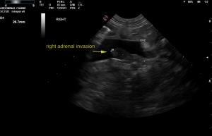
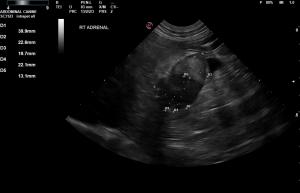
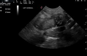
The right adrenal gland revealed early invasion into the vena cava that extended for approximately 2.8 cm. Hyperechoic nodular changes were noted throughout the right adrenal gland. The right adrenal gland itself measured 3.99 x 2.26 cm with capsular expansion and capsular escape into the vena cava. The right adrenal gland was significantly vascular. The left adrenal gland was also enlarged and hypervascular with regional phrenic and caval invasion measuring 1.35 cm. A mineralizing mass was deriving from the caudal pole and measured 2.7 cm.
The liver was riddled with multiple, mixed echogenic nodular changes. The largest of which measured 4.5 cm. There is a strong potential for metastatic disease. Passive congestion liver pattern. Gallbladder edema was noted.
The stomach was thickened with loss of curvilinear patterns. The gastric wall at the pyloric outflow measured 8.0 mm. The remainder of the intestine was unremarkable.
The pancreas was hypoechoic and irregular with coarse architecture and regional inflammation.
Regional inflammation was noted throughout the cranial abdomen.
Slight pericardial effusion was noted.


