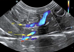
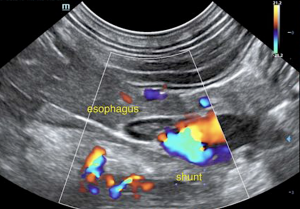
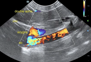
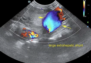
One of the biggest EHPSS SonoPath has ever seen; it's all about flow to the azygos. High end imaging performed by Diane McFadden, RVT, SDEP® certified clinical sonographer, and director of mobile operations and education for SonoPath. Expert interpretation by Eric Lindquist, DMV, DABVP, Cert. IVUSS, founder & CEO of SonoPath. Our SDEP hands-on wet labs as well as our SDEP Abdomen virtual lab incorporates training in imaging all shunts as part of the protocol; our intrahepatic and extrahepatic shunt posters are a fantastic reference with diagrams of each shunt as well as associated ultrasound images. Never miss that shunt!
DX
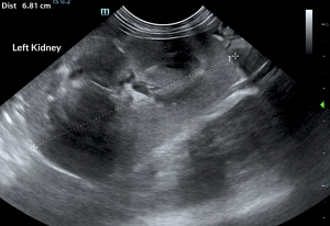
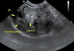
EHPSS: suspect splenoazygos shunt.
Outcome
Splenoazygos shunt is suspected. This is an extreme shunt fraction. An attempt at medical stabilization could be considered with potential surgical intervention. However, the size of the shunt is considerable and it is debatable on whether the liver would be able to handle the flow adequately without forming portal hypertension.Medical stabilization and CT for surgical planning and referral to Dr. Weisse at Animal Medical Center would be the preference for this case. Concurrent renal dysplasia is suspected; however, given the structural changes, the swelling of the kidneys can be justified by altered ammonia metabolism. Plasma transfusion as well as plasma expanders are likely in this patient’s best interest. After a prognosis of extremely guarded to poor, the patient was humanely euthanized.



Comments
Hepatic Support for Bile Acid Elevation +/-Hepatic Encephalopathy: Royal Canin Hepatic Support diet or Hills L/D, Metronidazole (7.5 mg/kg PO bid) over the next 14 days, Lactulose (Oral: 3.1-3.7 g/5 ml lactulose in a syrup base) long term to target 2-3 soft stools/day, with a high-quality protein supplement of minor amount of yogurt or cheddar cheese. Monitor bile acids, with attention paid to dropping albumin, BUN or cholesterol. SAMe and nutraceuticals as needed. Ursodiol (10-15 mg/kg p.o. q24h) can be considered as hepatoprotectant and to enhance bile flow. Zincserum level keep between 200—500 ug/dl. If deficient then Tx zinc acetate 1-3 mg/kg/day. Gastrointestinal protectants are recommended if the patient is anorexic.