DX
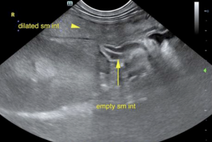
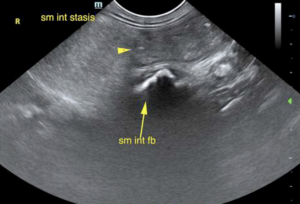
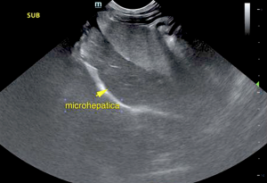
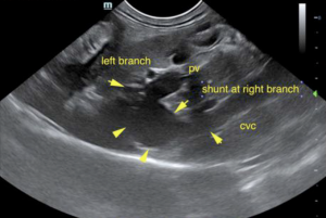
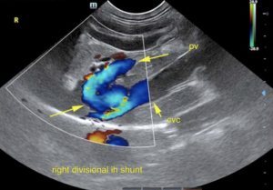
Distal small intestinal foreign body obstruction with concurrent intrahepatic right-divisional shunt.
Outcome
The immediate issue is the intestinal obstruction. The ability to metabolize anesthetic will be significantly decreased. Propofol and Isoflurane will likely be the best option for surgical intervention. Medical management for intrahepatic shunt is warranted post surgery with eventual vascular plug placement at Animal Medical Center *Dr. Weisse would be the most appropriate specialist to perform this procedure in the immediate region. Bile acid profile would be warranted, yet it is suspected to be significantly high. I recommend rapid surgery in this patient for the intestinal obstruction under the protocol recommended with followup treatment for the intrahepatic shunt. This is a congenital anomaly. The breeding line should be evaluated for intrahepatic shunting. There is a mild potential that the small intestinal foreign body could pass into the colon, yet was fully obstructed at the time of the sonogram. This appears to be a hard foreign body such as plastic or similar material.
The patient underwent surgery for the foreign body. An enterotomy was performed by Dr. Cattiny and Dr. Giammanco on 2/16/21. There were multiple, unidentifiable large foreign objects with strings attached that were removed from his intestines in addition to a sock. "The small intestines were noted to have large amount of necrosis in the descending duodenum and proximal jejunum. Adhesions present in the abdominal wall. Evidence of linear foreign body present in the pylorus/duodenum. Multiple foreign bodies present. Multiple large foreign objects removed with strings attached. The intestines were severely necrotic and ecchymotic." The patient had an uneventful recovery and experienced no complications. He was released from the hospital 2 days post-op. and owner reports he is doing excellent at home.


Comments
*Chick Weisse, VMD, DACVS can be found at the Animal Medical Center in New York City.