Contact Us or call 800-838-4268 for more information
Skip to main content

Splenocaval Liver Shunt In A 9-Month-Old Intact Male Shih Tzu: Our Case Of the Month August 2022

Patient Information

Age
9 Months
Gender
Male, Intact
Species
Canine

Images

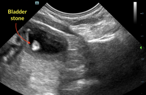
Bladder calculi.
Swollen, hypervascular left kidney.
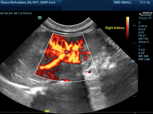
Swollen, hypervascular right kidney.
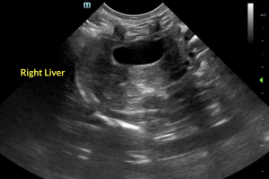
right liver
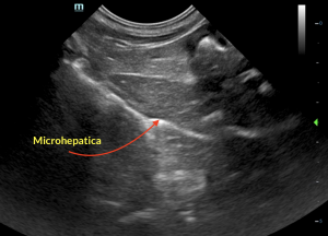
microhepatica
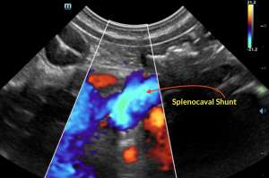
shunt entering vena cava
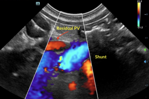
shunt entering vena cava, residual portal vein in red
shunt entering vena cava

A small breed puppy with a heart murmur, elevated pre & post bile acids, bladder calculi, and a small liver; shunt-hunt mode: ENGAGED. When evaluating the portal hilus what should the ratio of the 3 main vessels be to each other? The portal vein, caudal vena cava, and aorta in the portal hilus should be at a 1:1:1 ratio in a normal patient. SonoPath Mobile's Diane Mcfadden, BS, RVT, SDEP® certified clinical sonographer captured the high-end views needed to properly diagnose this little one's liver shunt. Detailed interpretation by Eric Lindquist, DMV, DABVP, Cert. IVUSS.

History

A 10 lb, 9-month-old intact male Shih Tzu puppy with a heart murmur presented for vomiting and lethargy following dietary indiscretion.

Image Interpretation

The urinary bladder revealed small calculi, the largest grouping of which measured 5.0 mm, nonobstructive at the time of the sonogram. The kidneys were swollen and hypervascular. The liver was subnormal in size with moderate to severe microhepatica. The gallbladder and common bile duct were unremarkable. The portal vein was subnormal in size. Extrahepatic portosystemic shunt present, width of approximately 8.0 mm, appeared to be deriving from the splenic vein juncture with the portal vein, decoursing dorsally and entering into the vena cava. The residual portal vein measured 4.0 mm. The shunt measured maximum 8.0 mm. The vena cava measured 7.0 mm. The aorta measured 5.0 mm at the portal hilus.

DX

Splenocaval shunt, bladder calculi, swollen kidneys, microhepatica.

Outcome

Clinical management with hepatic bile acid elevation support protocol was recommended until surgical intervention could occur for correction of splenocaval shunting. At last communication, the patient was having serial ultrasound rechecks to track the progression of the shunt.

Comments

If you need help with imaging liver shunts, see our online course "Sonographic Approach - Shunt Hunt 1 & 2. https://education.sonopath.com/ Detection of portosystemic shunts and the negative shunt hunt, utilizing the SDEP abdominal scanning protocol; exhaustive review of EHPSS and IHPSS including clinical approaches, medical and surgical management in this 2 part series.

Videos

Bladder stone.
Right liver.
Microhepatica.
Splenocaval shunt entering into the caudal vena cava and showing the residual portal vein.