Clinical Differential Diagnosis
Valvular disease, emerging CHF.
Image Interpretation
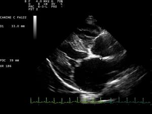
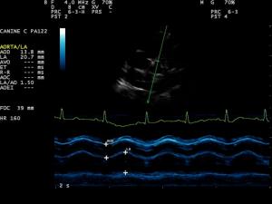
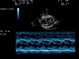
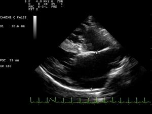
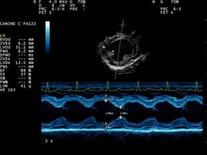
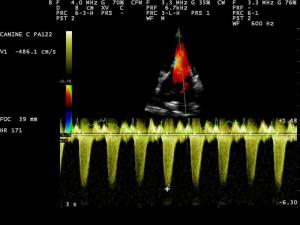
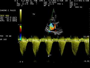
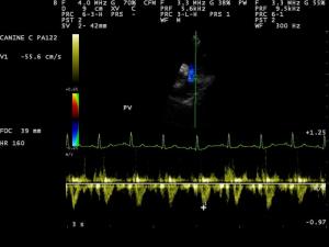
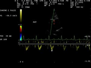
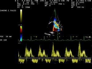
The left ventricle appears with mild to moderate volume overload. Systolic function is not impaired. The mitral valve leaflets, particularly the anterior one, are severely thickened; both leaflets are prolapsing. Severe mitral regurgitation is visible on color Doppler recordings. The left atrium is moderately enlarged. Mitral inflow profiles show a high E-wave amplitude and a short IVRT, indicating markedly increased left atrial pressures. The left ventricular outflow tact presents a normal morphology without evidence of fixed or dynamic obstruction. Left ventricular outflow is laminar; maximum outflow velocities do not exceed normal ranges. The right ventricle is mildly dilated. The tricuspid valve leaflets are markedly thickened and severe tricuspid regurgitation is noted on color Doppler clips. Systolic pressure gradients across the tricuspid valve are severely increased (~100 mm Hg). The right ventricular outflow tract, the pulmonic valve and the main pulmonary artery appear without morphological alterations. Systolic right ventricular outflow is laminar and there's no evidence of pulmonic stenosis. PW-profiles are clearly asymmetric which is indicative of pulmonary hypertension. A mild amount of pericardial effusion is seen. LV-dimensions IVSd 6.2 mm LVd 31.2 mm LVWd 6.2 mm IVSs10 mm LVs 13.5 mm LVWs 11 mm LA/AO 1.9.
Sonographic Differential Diagnosis
The findings are consistent with severe degenerative valve disease affecting both the mitral and tricuspid valve. Both atria are moderately enlarged. The left ventricle shows some amount of volume overload, the right ventricle is dilated. Pulmonary hypertension (PHT) is noted based on systolic TI gradients. The mild pericardial effusion in this case is likely caused by congestion even though other less likely causes (neoplasia, infectious are not completely ruled out). PHT is this case could be associated with increased downstream resistance (due to acute or chronic left sided heart failure). Other possible causes include: Thromboembolism (e.g. due to protein losing nephropathy or HAC), heart worm disease, primary airway disease, thoracic neoplasia, or pulmonary parenchymal disease.
DX
Stage C1 Valvular disease, Pulmonary Hypertension, Left and Right-sided Heart Failure.
Outcome
Re-evaluate the rads for the presence of signs of chronic left sided CHF, neoplasia or airway/pulmonary disease. Check for heart worm, protein losing nephropathy (UPC ratio), check for HAC and look for signs of airway disease. While results are pending, I would start with Pimobendan (0.25 mg/kg bid), an ACEI (e.g. Benazepril at 0.25 mg/kg bid) and Lasix (dosage dependent on the severity of ascites, e.g. 2 mg/kg bid), and do a follow up scan focused on PHT in 1-2 weeks.


Comments