
Making a great blood smear (or blood film as some call it) is an important skill for any veterinary professional, even if your hospital does not require differentials to be done on a regular basis. It’s a great way to impress your colleagues! It seems in this age of automated hematology analyzers, reading a differential is becoming a lost art. Plus it can be a lot of fun!
You never know when you may need to take a quick look to make sure platelet number is adequate, scan for blood parasites, or do a full differential if your analyzer is down (or you don’t have one!)
It all starts with a great blood smear!
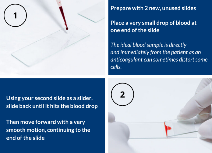
The smear itself should look very smooth with a seamless progression to what is called a “feathered edge”. This is the very end area of the smear and consists of a monolayer of cells. The monolayer will contain cells that are the easiest to identify and are the least distorted.
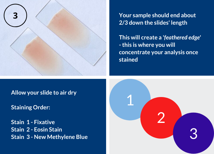
Too thin? (can occur with anemic patients) = Increase the angle of the slide you are using to smear with; otherwise your smear may run right off of your horizontal slide and you will not have a nice feathered edge, and your smear will be so thin you will have trouble finding enough cells.
Too thick? (can occur with dehydrated patients) = Decrease the angle of the slide to create a thinner smear; otherwise you will have cells piled on top of each other rather than a nice monolayer.
Really thick/takes up the entire slide? = Too large of a blood sample drop.

Try not to get impatient with yourself; it takes a lot of tries to finally perfect the motion and art of a great, diagnostic blood smear.
Practice, practice, practice!
