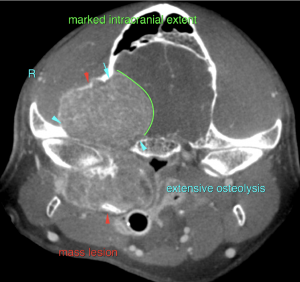
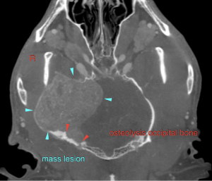
Head to Toe Diagnostic Efficiency™…. Clinical signs, a probe, a CT and expert interpretation means getting clinical answers and direction in same day turnaround. SonoPath has entered into functional on-site CT in cooperation with Blairstown Animal Hospital….and Dr Ken Leal.This patient had progressive clinical neuro signs with pathology visible in minutes…. What you can’t find with a probe, the CT will.
Many thanks to Dr. Sanam Sean Maniar, DVM and his team from Rockaway Animal Hospital for their exemplary care and management of this difficult case. We would like to also thank Sebastian Schaub, DVM, Dr. med. vet. DipECVDI for his interpretation of this case.
DX
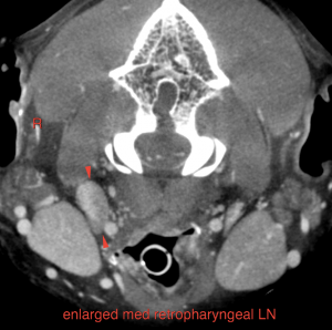
Biologically aggressive mass lesion of the right calvarium with aggressive osteolysis of the right temporal, occipital and sphenoid bone, extensive perforation of the cranial fossa and marked mass effect on the brain. Lymphadenomegaly of the right medial retropharyngeal lymph node.
Outcome
Main differential diagnoses are osteosarcoma, chondrosarcoma,multilobulated tumor of bone (chondrosarcoma rodens) and squamous cell carcinoma.
The lymph node changes are suggestive for metastatic spread.
Final diagnosis would require sampling of the primary tumor for histology and fine needle aspiration of the lymph node.
The lesion is not resectable. The long term prognosis is poor.
However, staging and palliative treatment such as tumor irradiation could be considered depending on the owner's wishes.


