Contact Us or call 800-838-4268 for more information
Skip to main content

Before and After Plavix

Patient Information

Age
10 Years
Gender
Male, Neutered
Species
Canine

Images

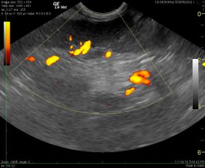
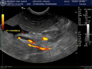
Power Doppler assessment of the portal vein demonstrates incomplete filling of the volume contracted vessel confirming portal vein thrombus. Spectral Doppler of the portal vein revealed lack of flow (not shown). Portal hypertension owing to portal vein thrombus was diagnosed.
Splenic vein thrombus is also identified with power Doppler. This is a common occurrence when the splenic vein thrombosis a cascade effect into the portal vein may occur. Yet ascites formation does not occur until the portal vein is adequately occluded to form portal hypertension.
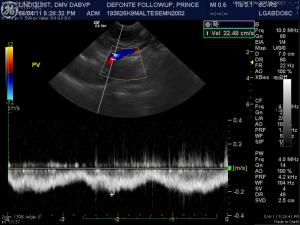
Portal vein velocity is measured with duplex Doppler. Normal velocity is present (18-25 cm/sec) indicating full resolution of portal hypertension.
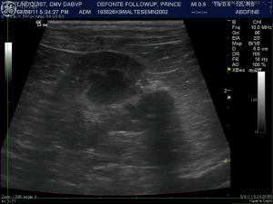
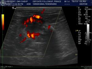
residual renal infarctions are present but renal values were never elevated in this patient indicating maintenance of adequate parenchymal function.
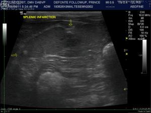
Permanent splenic infarction is also present with echogenic mineralization and capsular retraction. However, the splenic vasculature is patent and functional.

History

That dreadful swollen belly, life before and after Plavix, and a sneaky portal vein thrombus. Ever wonder how to resolve the mystery of that swollen abdomen in a 10-year-old hypercoagulable AIHA Maltese? Put a probe on it and see what’s really going on inside that belly. Working through the ascites case with the probe, Lindquist & a little help from his friends.

Case managed by Drs. Joe Powel and Paul Sedlacek & staff at Animal Clinic of Morris Plains, Morris Plains, New jersey, USA. Imaging by Dr. Eric Lindquist of SonoPath.com & New Jersey Mobile Associates.

Sonogram (Abdomen, 11/8/10, 3/4/11): “Prince”

History: A 10-year-old MN Maltese with recent history of profound anemia that had been diagnosed as primary autoimmune hemolytic anemia and had been treated with a blood transfusion, p rednisone and Atopica, was presented a few weeks later for anorexia, weight loss, and depression. Physical examination found the patient with generalized muscle wasting and a pot-bellied appearance. Abnormalities on CBC were mild anemia and leukocytosis. Doxycycline was added to the treatment to cover for infectious anemia pending an abdominal ultrasound.

Clinical Differential Diagnosis

(Lobetti): Drug effects – iatrogenic Cushing’s disease, gastric ulceration, steroid hepatopathy, infectious disease from immune suppression, pancreatitis Thrombo-embolic disease Pre-existing causes for IMHA – neoplasia, bacterial/fungal infections, abscess, granuloma

Sonographic Differential Diagnosis

(Lindquist DMV, DABVP): Portal vein thrombus with secondary portal hypertension and ascites. Splenic thrombosis. Renal infarctions.

Outcome

The patient was treated with Plavix and a plasma transfusion after the first sonogram was performed. At a recheck examination several weeks later, the patient was found to be doing very well on current therapies. CBC and blood chemistry were both within normal limits. The patient was recommended to continue with the current protocol of Plavix, Prednisone, and Atopica

Comments

Special thanks to Drs. Joe Powel and Paul Sedleck and staff at Animal Clinic of Morris Plains for their excellent and successful management of this complicated case.

Videos

Anechoic ascites is noted that was sampled and found to be transudate and not hemorrhage. The vena cava (4 cm depth traversing the diaphragm) is volume contracted ruling out passive congestion from the thorax (right chf, pericardial effusion, caudal thoracic mass..) as a cause of ascites. The liver parenchyma is uniform and no history supportive of diffuse liver disease was present. Hence portal hypertension owing to diffuse liver disease is unlikely. Echogenic lumen in the volume-contracted portal vein is visualized (3 cm depth).
Power Doppler assessment of the portal vein demonstrates incomplete filling of the volume contracted vessel confirming portal vein thrombus. Spectral Doppler of the portal vein revealed lack of flow (not shown). Portal hypertension owing to portal vein thrombus was diagnosed.
Splenic vein thrombus is also identified with power Doppler. This is a common occurrence when the splenic vein thrombosis a cascade effect into the portal vein may occur. Yet ascites formation does not occur until the portal vein is adequately occluded to form portal hypertension.
Renal infarctions are also visible and are likely of a prior date owing to the hyperechoic organization of the cortical insults. Power Doppler evidences the lack of blood flow to the infarcted regions. Plasma transfusion, Plavix and supportive fluid therapy and hospitalization allowed for discharge of this patient.
Portal vein (pv) volume is normal volume and power Doppler filling with a 1:1 ratio to the vena cava (cvc). No evidence of thrombus is present.
The liver is uniform with normal vasculature. No free fluid is noted.
Portal vein (pv) volume is normal volume and power Doppler filling with a 1:1 ratio to the vena cava (cvc). No evidence of thrombus is present.
residual renal infarctions are present but renal values were never elevated in this patient indicating maintenance of adequate parenchymal function.
Permanent splenic infarction is also present with echogenic mineralization and capsular retraction. However, the splenic vasculature is patent and functional.