Contact Us or call 800-838-4268 for more information
Skip to main content

Cardiac Hemangiosarcoma & Tamponade

Images

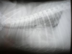
Radiograph: A single lateral thoracic radiograph reveals moderate right sided cardiomegaly with vascular congestion.
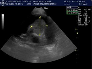
A 4 x 3.6 cm mass deriving from the heart base is noted in this short axis heart based view. The anechoic fluid surrounding the mass is owing to hemopericardium.
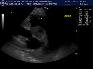
Oblique long axis view of the pericardial fluid and mass demonstrating the 16 g, 2-inch IV catheter placement into the pericardial sac for drainage. A suction unit is attached to an IV extension set for efficient drainage. The author utilizes a left lateral ventral 3-5 intercostal approach for pericardial drainage with local lidocaine block.

History

Cardiac Hemangiosarcoma & Tamponade. 6-month quality survival with adriamycin and pericardial drainage.

Lindquist (DABVP) & Nicolas (RDMS) of New Jersey Mobile, Ryan Epple DVM of Harmony AH, Philippsburgh, NJ, USA.

Sonogram (Cardiac): “Hawthorne”

History (Vasquez RVT, Lindquist DABVP): An 11-year-old MN Golden Retriever was presented for evaluation of lethargy and decreased appetite. Abnormalities on physical examination were lethargy and pulse deficits. Rectal temperature was normal. 4DX test showed a weak positive result for Anaplasma. Survey radiographs revealed cardiomegaly and suspicion of pleural effusion. Mild anemia was noted on CBC.

Clinical Differential Diagnosis

Clinical Differential Diagnosis (Lobetti DECVIM) Cardiac disease: Dilated cardiomyopathy Pericardial effusion: Idiopathic Neoplasia Pleural space disease: Neoplasia Diaphragmatic hernia Pyothorax Chylothorax Haemothorax Hypoproteinaemia Mediastinal obstruction/mass Lung disease: Parapneumonic effusion Lung lobe torsion

Image Interpretation

(Lindquist DABVP)

Sonographic Differential Diagnosis

(Lindquist DABVP): Heart based mass consistent with hemangiosarcoma and severe pericardial tamponade.

Sampling

US-guided thoracocentesis and pericardiocentesis were performed (Lindquist DABVP, Nicolas RDMS, Epple DVM).

Outcome

(Lindquist DABVP, Nicolas RDMS): Immediately after the pericardial effusion was drained, the patient was started on Doxorubicin therapy and walked away asymptomatic from the drainage procedure. He responded well to therapy and finished out five treatments of Doxorubicin. After more than four months the patient was reported to be doing well and clinically stable. Current therapy included omega-3 fatty acid supplement and NSAIDS. The patient survived with a good quality of life for 6 months at which time tumor regrowth to 3 times the original size, pericardial hemorrhage and tamponade caused excessive weakness compromising quality of life. Follow-up echocardiogram findings confirmed this suspicion. The owners were very content with the extra 6 months of quality time even though empirical chemotherapeutic intervention was the mainstay of treatment.

Comments

Special thanks to: Dr. Ryan Epple and staff at Harmony Animal Hospital, Phillipsburgh, NJ, USA, as well as the Kane family for their unsurpassed compassion and management of Hawthorne's pathology.

Videos

4-chamber long axis echocardiogram reveals severe pericardial effusion and tamponade. A 5 cm hypoechoic irregular mass deriving from the right atrial and right ventricular free wall junction strongly suggestive for hemangiosarcoma.
Short axis left ventricular echocardiogram demonstrates volume contraction and pseudohypertrophy of the left ventricle and severe pericardial tamponade; which is responsible for the generalized cardiomegaly on the radiograph. Paradoxically the internal volume of the heart is significantly compromised.
5-chamber long axis view of the heart after drainage of 250 cc of frank blood from the pericardial sac. The near field anechoic fluid represents the residual hemopericardium; which is at this point minimal and does not impede atrial and ventricular filling.