Contact Us or call 800-838-4268 for more information
Skip to main content

Collapse and Cyanosis

Patient Information

Age
11 Years
Gender
Male, Neutered
Species
Canine

Images

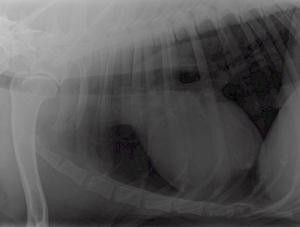
Survey radiographs showed a mildly enlarged cardiac silhouette (VHS 11), prominent right ventricle, normal left atrium, and a normal lung pattern.
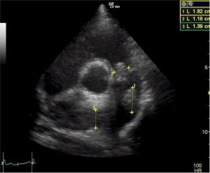
the masses can be clearly measured. Given the solid structural organization consistent with tissue, simple thrombosis is unlikely.
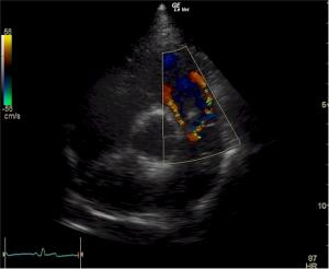
On CDI it can be seen very clearly that the masses are causing obstruction to flow and seem to be somehow confluent on these clips.
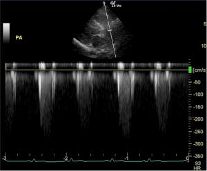
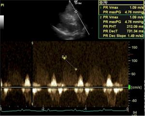
Flow velocity across the RVOT and PV is increased (3.2 m/s), but v-max of the pulmonary insufficiency noted is normal excluding pulmonary hypertension.

History

It’s a clot! No it’s a thrombus! Or… maybe it’s a mass… What are those things messing with Tomie Timon’s Doppler in the pulmonic outflow tract in this echocardiogram?

Tomie Timon RDMS of Eugene, Oregon, shows us “what does this” with Dr. Peter Modler’s help from sunny AustriaJ. Oh yes the world is truly flat Mr. Friedman when veterinary diagnoses come from half way across the planet by means of an Oregon-Austrian connection.

Imaging by Tomie Timon RDMS, Animal Sounds Mobile Veterinary Ultrasound Service, Eugene, Oregon.

History: An 11-year-old NM Golden retriever was presented for evaluation of two episodes of collapse and cyanosis. Additional history was that the patient is more lethargic than usual. Cardiac auscultation did not reveal any abnormalities and blood pressure was normal.

Clinical Differential Diagnosis

Pericardial effusion Cardiomyopathy – hypertrophic, dilated Myocarditis Cardiac neoplasia Pulmonic thrombo-embolic disease Metabolic disease – anemia, met-hemoglobinemia

Sonographic Differential Diagnosis

(Modler): The most remarkable abnormality is the presence of a mass or masses within the pulmonary artery and attached to the pulmonic valve. All masses have the same echogenicity and cause obstruction to flow which the consequence of right ventricular hypertrophy. Even though it cannot be ruled out that these masses are thrombi (but not septic thrombi because there is no history of fever) I would think that this is neoplasia (thrombosis usually causes pulmonary hypertension). One case of pulmonary artery leiomyosarcoma is mentioned in the veterinary literature, more can be found in human literature. Obtaining a sample via catheter (snare) would be ideal If this is not possible then the only option would be r ight heart surgery (no cardioplegia necessary). And I would recommend administration of Plavix or aspirin in this patient as a palliative measure and recheck sonogram weekly if no sampling is possible.

Sampling

Post mortem biopsy revealed marked lung congestion, right ventricular hypertrophy, and grade 3 fibrosarcoma of the pulmonary valve with attached thrombus.

Comments

Special thanks to Dr. Nancy Johnson at The Veterinary Hospital, Eugene, OR for managing this case; and to Dr. Mark McConnell at Emergency Veterinary Hospital, Eugene for necropsy.

Videos

There is a mass (1.18 cm) attached to and moving with the pulmonary valve. Another mass (1.92 cm) is seen along the cranio dorsal border of the PA. The right pulmonary artery branch is obviously filled with another homogenous mass.
On CDI it can be seen very clearly that the masses are causing obstruction to flow and seem to be somehow confluent on these clips.