Contact Us or call 800-838-4268 for more information
Skip to main content

Cushing's Disease

Patient Information

Age
13 Years
Gender
Female, Spayed
Species
Feline

Images

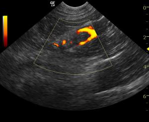
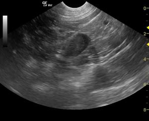
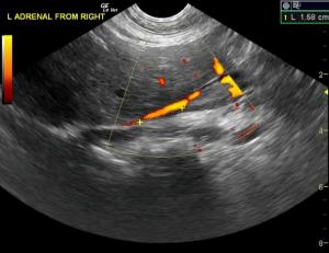
There is a left adrenal mass in this patient that is invading the phrenicoabdominal vein.
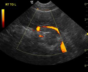
The echogenic change in the caudal vena cava (CVC) is occupying approximately 80% of the vessel and occupies a length of 4.1cm. Power Doppler demonstrates the flow around the invasive tumor or thrombus.
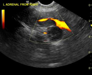
Scanning the left adrenal from the right side allows for better visualization of the vena cava and the entrance of the phrenic vein into the CVC where the tumor is invading with potential attached thrombus. It is difficult to ascertain which part is tumor and which part is thrombus as they have the same echogenicity.
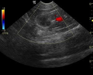
Follow-up sonogram 6 months form the original diagnosis utilizing similar approaches to those in images 1-5 reveal further invasion/thrombus progression .

History

Stanley has Cushing's disease. Is Stanley a Beagle? A Dachshund? A Yorkie? No, Stanley is a DSH house cat and take a look what his adrenal was doing and how the disease progressed but still allowed Stanley to have some quality time. "Give adrenals a chance."

Imaging and clinical interpretation by Eric Lindquist DMV, DABVP.

History: A 13-year-old FS DSH cat was presented for the evaluation of polyuria and polydipsia of several months’ duration. The serum biochemical profile revealed marked hyperglycemia. The CBC showed a low RBC count, lymphopenia, and a monocytosis. The TT4 and free T4 were within normal limits. A fructosamine level was high. The urinalysis showed glucosuria, and hematuria. The urine culture was negative for bacterial growth. A fructosamine level was repeated a few months later and still showed an elevated fructosamine. Insulin-like Growth Factor 1 was normal, and a diagnosis of acromegaly was considered unlikely. A recheck of the blood work revealed hyperglycemia, hyperphosphatemia, hypokalemia and hyperlipasemia. The CBC showed elevations of the MCH and MCHC, as well as thrombocytopenia. The results of an ACTH stimulation were suggestive of hyperadrenocorticism. The urine had a turbid appearance on the urinalysis and showed proteinuria, hematuria, and elevated RBCs microscopically. The urine culture was negative. Vitamin B12, folate concentrations were performed and the vitamin B12 was within the normal reference range, while the folate was increased. High fTLI and a high PLI were also present. Fructosamine levels were now within normal limits.

Clinical Differential Diagnosis

Diabetes mellitus in conjunction with pancreatitis, hypoaldosteronism secondary to an adrenal mass, such as an adenocarcinoma or adenoma.

Image Interpretation

The adrenal gland in this patient has grown from size from 2 – 2.5cm. Caval invasion through phrenic vein has occurred at increased distance of 7cm and approximately 1.5cm from the diaphragmatic inlet in the vena cava.”

Sampling

None taken

Outcome

The patient was referred for an abdominal CT scan. A heterogeneous, hypodense soft tissue mass obliterated the normal architecture of the left adrenal gland. There was also invasion of the caudal vena cava at the junction of the left renal vein and the middle of the mass. The right adrenal gland was not visualized. A focal, hypodense nodule was present in the caudate liver lobe. The cat was suspected to have a neoplasm affecting the left adrenal gland with metastases to the caudal vena cava, as well as possible left renal vein involvement. Presence of metastases to the liver could not be ruled out as not all of the liver was included in the study. Medical management was determined to be the best option in this case, as the patient was not considered a good candidate for surgery.

Videos

Video of the left adrenal invasive process enhanced by color flow Doppler.
Follow-up sonogram 6 months form the original diagnosis utilizing similar approaches to those in images 1-5 reveal further invasion/thrombus progression .