How many kidneys does a cat really need? Extra parts, unusual parts, and not enough parts in 3 cats with “wacked” kidneys and a little pancreas comprise the SonoPath cases of the month. Imaging by Casey DVM DABVP, Parkinson RDMS, & Lindquist DMV, DABVP.
So perusing through the SonoPath archive while working on the book, (yes we still working on it and are making progress) I came across these 3 peculiar feline kidney cases. Two cases mother-nature must have read my wine recommendations in the lifestyle section of SonoPath.com and took the recommendations a bit too much to heart when firing up the urology genetics on these first 2 kitties of 3 in this series. Or perhaps she was suffering from the post-prandial tide after too much pasta with my Bolognese sauce and she lost her concentration?
Now what other veterinary site can you find a myriad of sonographic pathology along with wine and food recommendations? You have to love the Internet as there truly are few rules as long as the material is done with taste, so to speak.
In addition to the 2 congenitally deformed kidney cats, there is the third cat in this series that has 3 kidneys owing a renal transplant where the allograft kidney is basically slung (with technical precision) onto the body wall cranial to the urinary bladder. However, this cat was being treated well for pyelonephritis but was significantly painful and lethargic for concurrent pancreatitis. This is a scenario I have observed very often in cats that have only mild/moderate degenerative or pathological renal changes and concurrent pancreatitis seems to be causing them to circle the drain with “renal failure” parameters. But these patients often respond remarkably to “pancreatitis” therapy and pain management in addition to the typical IV renal protocol. Have you observed the same? Questions like this will be addressed in the new SonoPath image/video up-loadable forum coming in the Fall along with the clinical and pathology search on SonoPath.com.
Moreover, for those of you coming to the IVUSS conference in Bologna, Italy September 12-16, I am looking forward to meeting you there.
Take a look at these 3 sonographic scenarios with the help of my good friends and colleagues Dr. Doug Casey of English Bay Ultrasound, Vancouver, BC, Canada, and Andi Parkinson RDMS of Intrapet Imaging, Baltimore, MD, USA.
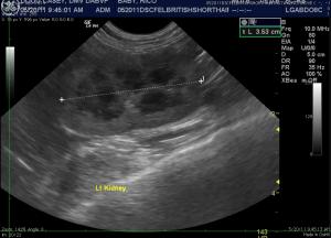
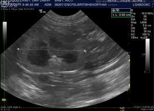
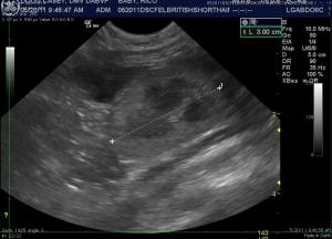
Case 1: Baby is a 3-year old British Short Haired cat that was imaged for non-urinary reasons (Diarrhea). The third kidney was an incidental finding and certainly made Dr. Doug Casey have a second and third look at things. A small calculus is present in the pelvis of the right kidney. The third vestigial kidney is mildly subnormal in size (2.5 cm) and located dorsal to the urinary bladder.
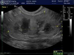
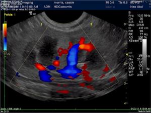
Case 2: Cassie is a 2-year-old Domestic Short Haired cat that presented for hematuria and mildly elevated creatinine. The kidneys (2 total) appeared fused at the renal pelvis with a dual vascular supply. Bilateral renal pelvic dilation was noted and chronic urinary tract infection is likely or the pelvic dilation may be part of the primary dysplastic structure. Cortical vascularity appeared normal in both kidneys. The one kidney measured 3.28 cm and the other 3.4 cm individually. However, the right and left renal fusion together in long axis measured 5.2 cm.
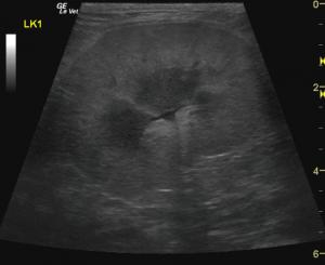
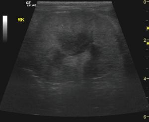
Case 3: Boots is a 10.5 year-old Domestic Short haired cat that presented for lethargy and anorexia persistent for 1-week post renal transplant surgery. The patient was treated with immune suppressive medications and had persistent but strongly regenerative anemia and culture + for urinary tract infection as well as elevated BUN and creatinine levels. The original left and right kidneys present sub-vascular echogenic cortices consistent with interstitial nephrosis. The allograft kidney is surgically tethered to the body wall cranial to the urinary bladder. Renal pelvic dilation is also present with ill-defined pelvic architecture. However, cortical vascularity is strong, evidenced on power Doppler imaging. A slight amount of adjacent free fluid is noted that resolved on follow-up ultrasound 5 days later. The pancreas is distinctly hypoechoic and irregular with a + Murphy sign upon imaging this region. Enlargement of the pancreatic parenchyma, irregular contour, ill-defined surrounding hyperechoic fat, and pain upon imaging are typical parameters for feline pancreatitis. The patient responded to medical therapy and pain management over the following 2 weeks and was discharged with a persistently regenerative anemia.


Comments